• 文献检索
  • 文档翻译
  • 深度研究
  • 学术资讯
  • Suppr Zotero 插件Zotero 插件
  • 邀请有礼
  • 套餐&价格
  • 历史记录
应用&插件
Suppr Zotero 插件Zotero 插件浏览器插件Mac 客户端Windows 客户端微信小程序
定价
高级版会员购买积分包购买API积分包
服务
文献检索文档翻译深度研究API 文档MCP 服务
关于我们
关于 Suppr公司介绍联系我们用户协议隐私条款
关注我们

Suppr 超能文献

核心技术专利:CN118964589B侵权必究
粤ICP备2023148730 号-1Suppr @ 2026

文献检索

告别复杂PubMed语法,用中文像聊天一样搜索,搜遍4000万医学文献。AI智能推荐,让科研检索更轻松。

立即免费搜索

文件翻译

保留排版,准确专业,支持PDF/Word/PPT等文件格式,支持 12+语言互译。

免费翻译文档

深度研究

AI帮你快速写综述,25分钟生成高质量综述,智能提取关键信息,辅助科研写作。

立即免费体验

在一名患有脐膨出且接受分期手术的婴儿中,采用内镜下成分分离技术成功闭合巨大腹壁缺损。

Successful closure of a large abdominal wall defect using endoscopic component separation technique in an infant with a giant ventral hernia after staged surgery for omphalocele.

作者信息

Toma Miki, Yanai Toshihiro, Yoshida Shiho

机构信息

Department of Pediatric Surgery, Ibaraki Children's Hospital, 3-3-1, Futabadai, Mito, Ibaraki, 311-4145, Japan.

出版信息

Surg Case Rep. 2021 Jan 6;7(1):4. doi: 10.1186/s40792-020-01087-2.

DOI:10.1186/s40792-020-01087-2
PMID:33409717
原文链接:https://pmc.ncbi.nlm.nih.gov/articles/PMC7788121/
Abstract

BACKGROUND

The management of large abdominal wall defects, such as omphalocele or gastroschisis, remains a challenge for pediatric surgeons. Though several techniques have been described to repair those conditions, there is no procedure considered to be the standard worldwide. We report an infant girl with a giant ventral hernia after staged surgery for omphalocele in whom delayed closure of a large abdominal wall defect was performed successfully using "endoscopic component separation technique (ECST)" without serious complications.

CASE PRESENTATION

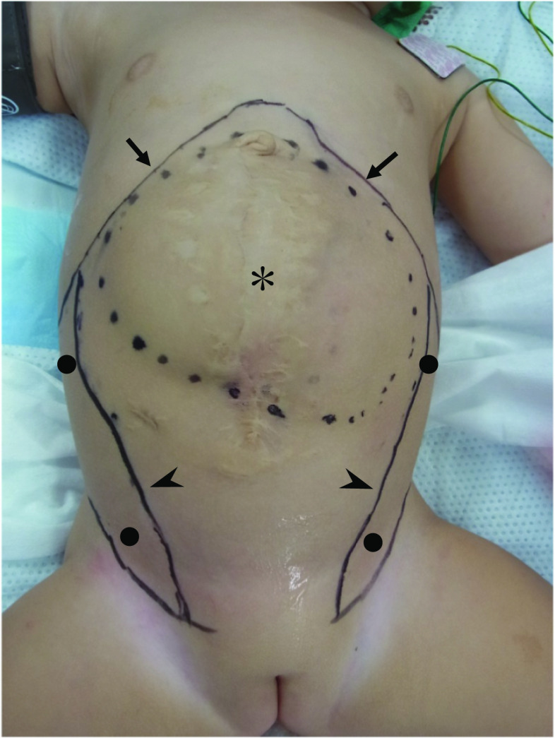
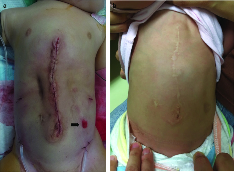
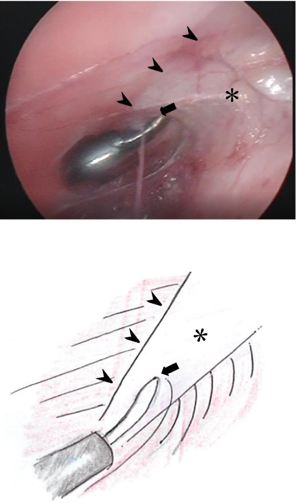
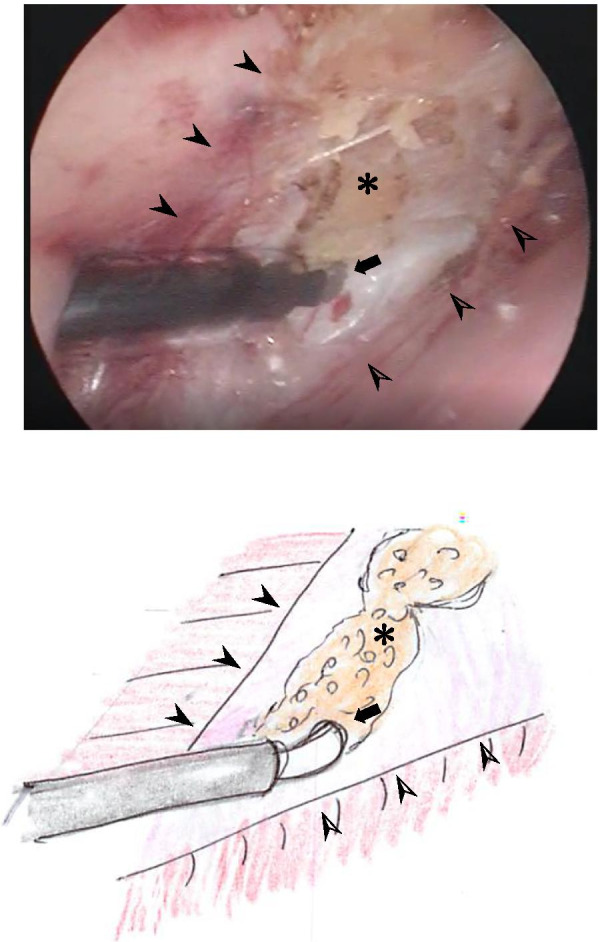
A baby girl was admitted to our hospital because of a giant omphalocele, which had been prenatally diagnosed. The omphalocele was supraumbilical and included the entire liver. After staged surgery, a large abdominal wall defect was closed by skin, creating a giant ventral hernia. We performed endoscopic separation component technique (ECST) for the closure of her abdominal wall defect when she was 11 months of age. ECST was initiated with placement of a 5.0-mm port just above the inguinal ligament and under the external oblique muscle. The space between the external and internal oblique muscles was created by the insufflation pressure, and a second 5.0-mm port was placed at 1.0 cm below the inferior edge of the rib into the space. As the further dissection was carried, the aponeurosis of the external oblique muscle was identified as a white line, running vertically from the epigastrium to inguinal ligament. It was transected longitudinally using electrocautery over its full length. The same procedure was performed on the contralateral side and the abdominal wall was successfully closed. Postoperative course was uneventful.

CONCLUSIONS

The technique of ECST, described here, is simple and safe for infants, and the cosmetic result is satisfactory.

摘要

背景

大型腹壁缺损,如脐膨出或腹裂的处理,对小儿外科医生来说仍然是一项挑战。尽管已有多种技术用于修复这些病症,但尚无一种被认为是全球标准的手术方法。我们报告了一名婴儿女孩,她在接受脐膨出分期手术后出现巨大腹侧疝,采用“内镜下成分分离技术(ECST)”成功进行了大型腹壁缺损的延迟关闭,且未出现严重并发症。

病例介绍

一名因巨大脐膨出入院的女婴,该脐膨出在产前已被诊断。脐膨出位于脐上,包含整个肝脏。分期手术后,腹壁缺损用皮肤关闭,形成巨大腹侧疝。当她11个月大时,我们采用内镜分离成分技术(ECST)来关闭其腹壁缺损。ECST开始时,在腹股沟韧带上方、腹外斜肌下方置入一个5.0毫米的端口。通过气腹压力在腹外斜肌和腹内斜肌之间形成空间,并在肋骨下缘下方1.0厘米处向该空间置入第二个5.0毫米的端口。随着进一步的解剖,腹外斜肌腱膜被识别为一条从腹上区垂直延伸至腹股沟韧带的白线。使用电灼术在其全长范围内纵向切断。对侧进行相同操作,腹壁成功关闭。术后过程顺利。

结论

本文所述的ECST技术对婴儿来说简单且安全,美容效果令人满意。

https://cdn.ncbi.nlm.nih.gov/pmc/blobs/7ead/7788121/cf6444832d8c/40792_2020_1087_Fig6_HTML.jpg
https://cdn.ncbi.nlm.nih.gov/pmc/blobs/7ead/7788121/d945cbc1ffb2/40792_2020_1087_Fig1_HTML.jpg
https://cdn.ncbi.nlm.nih.gov/pmc/blobs/7ead/7788121/a3381709b69f/40792_2020_1087_Fig2_HTML.jpg
https://cdn.ncbi.nlm.nih.gov/pmc/blobs/7ead/7788121/7a8a59eb7e94/40792_2020_1087_Fig3_HTML.jpg
https://cdn.ncbi.nlm.nih.gov/pmc/blobs/7ead/7788121/d867c711a287/40792_2020_1087_Fig4_HTML.jpg
https://cdn.ncbi.nlm.nih.gov/pmc/blobs/7ead/7788121/de87ebee49c2/40792_2020_1087_Fig5_HTML.jpg
https://cdn.ncbi.nlm.nih.gov/pmc/blobs/7ead/7788121/cf6444832d8c/40792_2020_1087_Fig6_HTML.jpg

相似文献

1
Successful closure of a large abdominal wall defect using endoscopic component separation technique in an infant with a giant ventral hernia after staged surgery for omphalocele.在一名患有脐膨出且接受分期手术的婴儿中,采用内镜下成分分离技术成功闭合巨大腹壁缺损。
Surg Case Rep. 2021 Jan 6;7(1):4. doi: 10.1186/s40792-020-01087-2.
2
Abdominal wall defect repair with component separation technique for giant omphalocele with previous relaxing incisions on the abdominal skin.采用腹壁组织分离技术修复巨大脐膨出且既往腹部皮肤有松弛切口的腹壁缺损。
Surg Case Rep. 2023 Jun 7;9(1):99. doi: 10.1186/s40792-023-01679-8.
3
Inguinal single-port approach of endoscopic component separation for abdominal wall defects: A case series.腹股沟单孔法内镜下腹壁缺损组织分离术:病例系列
Ann Med Surg (Lond). 2022 Sep 9;82:104611. doi: 10.1016/j.amsu.2022.104611. eCollection 2022 Oct.
4
Evolution of Endoscopic Anterior Component Separation to a Precostal Access with a New Cylindrical Balloon Trocar.
J Laparoendosc Adv Surg Tech A. 2018 Jun;28(6):730-735. doi: 10.1089/lap.2017.0480. Epub 2017 Oct 20.
5
Single port component separation: endoscopic external oblique release for complex ventral hernia repair.单孔组件分离:内镜下腹外斜肌松解在复杂腹疝修补中的应用。
Surg Endosc. 2018 May;32(5):2474-2479. doi: 10.1007/s00464-017-5949-3. Epub 2017 Dec 20.
6
Components separation technique for large abdominal wall defect.巨大腹壁缺损的成分分离技术
J Korean Surg Soc. 2011 Jun;80 Suppl 1(Suppl 1):S63-6. doi: 10.4174/jkss.2011.80.Suppl1.S63. Epub 2011 Jun 17.
7
Tissue Expander-Assisted Component Separation for Pediatric Abdominal Wall Reconstruction.组织扩张器辅助的小儿腹壁重建的组件分离。
Ann Plast Surg. 2022 May 1;88(4 Suppl 4):S320-S324. doi: 10.1097/SAP.0000000000003138.
8
Preoperative Botulinum toxin A enabling defect closure and laparoscopic repair of complex ventral hernia.术前注射肉毒毒素 A 以实现缺损闭合,并进行腹腔镜修复复杂的腹疝。
Surg Endosc. 2018 Feb;32(2):831-839. doi: 10.1007/s00464-017-5750-3. Epub 2017 Jul 21.
9
Component separation for complex congenital abdominal wall defects: not just for adults anymore.复杂先天性腹壁缺损的分离技术:不再仅仅适用于成人。
J Pediatr Surg. 2013 Dec;48(12):2525-9. doi: 10.1016/j.jpedsurg.2013.05.067.
10
Component Separation Technique for Repair of Massive Abdominal Wall Defects at a Pediatric Hospital.一家儿科医院用于修复巨大腹壁缺损的组件分离技术
Ann Plast Surg. 2016 Nov;77(5):555-559. doi: 10.1097/SAP.0000000000000652.

引用本文的文献

1
Giant omphalocele: A novel approach for primary repair in the neonatal period using botulinum toxin.巨大脐膨出:新生儿期应用肉毒毒素行一期修复的新方法。
Rev Col Bras Cir. 2023 Nov 20;50:e20233582. doi: 10.1590/0100-6991e-20233582-en. eCollection 2023.

本文引用的文献

1
How to perform the endoscopically assisted components separation technique (ECST) for large ventral hernia repair.如何进行内镜辅助下的腹壁大切口疝修补术的成分分离技术(ECST)。
Hernia. 2016 Jun;20(3):441-7. doi: 10.1007/s10029-016-1485-7. Epub 2016 Apr 1.
2
Large and complex ventral hernia repair using "components separation technique" without mesh results in a high recurrence rate.采用“成分分离技术”且不使用补片进行大型复杂腹侧疝修补术会导致较高的复发率。
Am J Surg. 2015 Jan;209(1):170-9. doi: 10.1016/j.amjsurg.2014.02.013. Epub 2014 Apr 28.
3
Component separation for complex congenital abdominal wall defects: not just for adults anymore.
复杂先天性腹壁缺损的分离技术:不再仅仅适用于成人。
J Pediatr Surg. 2013 Dec;48(12):2525-9. doi: 10.1016/j.jpedsurg.2013.05.067.
4
The component separation technique for hernia repair: a comparison of open and endoscopic techniques.疝修补的成分分离技术:开放技术与内镜技术的比较
Am Surg. 2011 Jul;77(7):839-43.
5
Endoscopic versus open component separation in complex abdominal wall reconstruction.内镜与开放型组件分离术在复杂腹壁重建中的应用比较。
Am J Surg. 2010 Mar;199(3):342-6; discussion 346-7. doi: 10.1016/j.amjsurg.2009.09.015.
6
Closure of giant omphaloceles by the abdominal wall component separation technique in infants.采用腹壁成分分离技术闭合婴儿巨大脐膨出。
J Pediatr Surg. 2008 Jan;43(1):246-50. doi: 10.1016/j.jpedsurg.2007.09.051.
7
Laparoscopic component separation in the single-stage treatment of infected abdominal wall prosthetic removal.腹腔镜下成分分离术在感染性腹壁假体取出的单阶段治疗中的应用
Hernia. 2007 Oct;11(5):435-40. doi: 10.1007/s10029-007-0255-y. Epub 2007 Jul 24.
8
Secondary closure of a giant omphalocele by translation of the muscular layers: a new method.
Pediatr Surg Int. 2005 May;21(5):373-6. doi: 10.1007/s00383-005-1387-8. Epub 2005 Apr 1.
9
Endoscopically assisted "components separation" for closure of abdominal wall defects.内镜辅助“成分分离”技术用于腹壁缺损的闭合
Plast Reconstr Surg. 2000 Feb;105(2):720-9; quiz 730. doi: 10.1097/00006534-200002000-00039.
10
"Components separation" method for closure of abdominal-wall defects: an anatomic and clinical study.用于腹壁缺损闭合的“成分分离”方法:一项解剖学与临床研究
Plast Reconstr Surg. 1990 Sep;86(3):519-26. doi: 10.1097/00006534-199009000-00023.