Suppr超能文献

产前阿片类药物暴露和吸烟对胎儿 MRI 中胎儿大脑发育和形态计量学的初步研究。

Pilot study of fetal brain development and morphometry in prenatal opioid exposure and smoking on fetal MRI.

机构信息

Department of Radiology and Imaging Sciences, Indiana University School of Medicine, Indianapolis, IN, United States.

Department of Radiology and Imaging Sciences, Indiana University School of Medicine, Indianapolis, IN, United States.

出版信息

J Neuroradiol. 2022 Jan;49(1):53-58. doi: 10.1016/j.neurad.2020.12.004. Epub 2021 Jan 5.

Abstract

PURPOSE

The purpose of this study was to assess for any differences in brain maturation, structure and morphometry in fetuses exposed to opioids in utero, compared to non-opioid exposed fetuses on fetal MRI.

METHODS

We performed a prospective study in pregnant women using opioids and healthy pregnant women without prenatal opioid use. We evaluated brain maturation, structure, and morphometry on second or third trimester fetal MRI and assessed group differences.

RESULTS

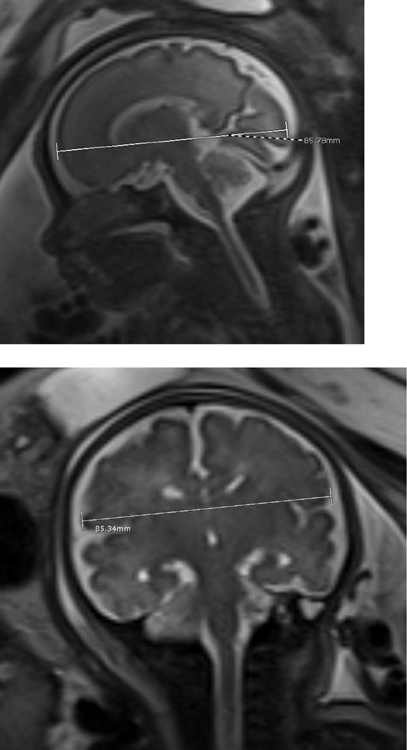
28 pregnant women were enrolled, 12 with opioid exposure (average gestational age 33.67, range 28-39 w), 9 of whom also smoked, and 16 without opioid exposure (average gestational age 32.53, range 27-38 w). There was a significant difference in the anteroposterior diameter of the fetal cerebellar vermis in the opioid exposed fetuses compared to non-opioid exposed fetuses (p = 0.004). There were no significant differences in brain biparietal diameter, fronto-occipital diameter, transverse cerebellar diameter and anteroposterior dimension of the pons in opioid exposed fetuses compared to non-opioid exposed fetuses. There were no abnormalities in brain maturation and no major brain structural abnormalities in the opioid exposed fetuses.

CONCLUSION

Smaller fetal anteroposterior cerebellar vermian dimension was associated with in utero opioid exposure. There were no abnormalities in brain maturation or major structural abnormalities in fetuses exposed to opioids.

摘要

目的

本研究旨在评估胎儿在宫内暴露于阿片类药物与非阿片类药物暴露胎儿之间在大脑成熟度、结构和形态测量方面的差异,方法:我们对使用阿片类药物的孕妇和未使用产前阿片类药物的健康孕妇进行了前瞻性研究。我们在胎儿 MRI 上评估了胎儿在第二或第三个三个月的大脑成熟度、结构和形态测量,并评估了组间差异。结果:共纳入 28 名孕妇,12 名暴露于阿片类药物(平均孕龄 33.67,范围 28-39 周),其中 9 名还吸烟,16 名未暴露于阿片类药物(平均孕龄 32.53,范围 27-38 周)。与非阿片类药物暴露胎儿相比,阿片类药物暴露胎儿的小脑蚓部前后径有显著差异(p=0.004)。与非阿片类药物暴露胎儿相比,阿片类药物暴露胎儿的大脑双顶径、额枕径、小脑横径和桥前前后径无显著差异。阿片类药物暴露胎儿的大脑成熟无异常,也无主要大脑结构异常。结论:小脑蚓部前后径较小与胎儿宫内阿片类药物暴露有关。阿片类药物暴露胎儿的大脑成熟或主要结构异常。

相似文献

1
Pilot study of fetal brain development and morphometry in prenatal opioid exposure and smoking on fetal MRI.
J Neuroradiol. 2022 Jan;49(1):53-58. doi: 10.1016/j.neurad.2020.12.004. Epub 2021 Jan 5.
2
MRI Findings in Third-Trimester Opioid-Exposed Fetuses, With Focus on Brain Measurements: A Prospective Multicenter Case-Control Study.
AJR Am J Roentgenol. 2023 Mar;220(3):418-427. doi: 10.2214/AJR.22.28357. Epub 2022 Sep 28.
4
Fetal brain morphometry on prenatal magnetic resonance imaging in congenital diaphragmatic hernia.
Pediatr Radiol. 2019 Feb;49(2):217-223. doi: 10.1007/s00247-018-4272-z. Epub 2018 Oct 6.
6
Can prenatal ultrasound detect the effects of in-utero alcohol exposure? A pilot study.
Ultrasound Obstet Gynecol. 2009 Jun;33(6):683-9. doi: 10.1002/uog.6379.
7
Fetal assessment in buprenorphine-maintained women using fetal magnetoencephalography: a pilot study.
Addiction. 2018 Oct;113(10):1895-1904. doi: 10.1111/add.14266. Epub 2018 Jun 13.
8
Brain cortical assessment by MRI in fetuses with left congenital diaphragmatic hernia.
Prenat Diagn. 2023 Jul;43(8):1002-1007. doi: 10.1002/pd.6368. Epub 2023 May 22.
9
Normal human brainstem development in vivo: a quantitative fetal MRI study.
Ultrasound Obstet Gynecol. 2021 Aug;58(2):254-263. doi: 10.1002/uog.22162. Epub 2021 Jul 6.
10
DTI of Opioid-Exposed Fetuses Using ComBat Harmonization: A Bi-Institutional Study.
AJNR Am J Neuroradiol. 2023 Sep;44(9):1084-1089. doi: 10.3174/ajnr.A7951. Epub 2023 Aug 10.

引用本文的文献

1
Antenatal Opioid Exposure and Global and Regional Brain Volumes in Newborns.
JAMA Pediatr. 2025 Apr 7. doi: 10.1001/jamapediatrics.2025.0277.
2
Children had smaller brain volumes and cortical surface areas after prenatal opioid maintenance therapy exposure.
Acta Paediatr. 2025 Feb;114(2):398-409. doi: 10.1111/apa.17448. Epub 2024 Oct 8.
3
Neuroimaging of opioid effects in humans across conditions of acute administration, chronic pain therapy, and opioid use disorder.
Trends Neurosci. 2024 Jun;47(6):418-431. doi: 10.1016/j.tins.2024.04.005. Epub 2024 May 17.
5
Prenatal Opioid Exposure and Risk for Adverse Brain and Motor Outcomes in Infants Born Premature.
J Pediatr. 2024 Apr;267:113908. doi: 10.1016/j.jpeds.2024.113908. Epub 2024 Jan 12.
6
Ultrasound evaluation of brain parenchyma in preterm infants with prenatal opioid exposure.
J Perinatol. 2024 Aug;44(8):1119-1124. doi: 10.1038/s41372-023-01804-3. Epub 2023 Oct 20.
7
Effects of medications for opioid use disorder (MOUD) on fetal brain and cranial measurements.
Neurotoxicol Teratol. 2023 May-Jun;97:107177. doi: 10.1016/j.ntt.2023.107177. Epub 2023 Apr 19.
8
Placental volume in pregnant women with opioid use: prenatal MRI assessment.
J Matern Fetal Neonatal Med. 2023 Dec;36(1):2157256. doi: 10.1080/14767058.2022.2157256.
10
Brain structural connectome in neonates with prenatal opioid exposure.
Front Neurosci. 2022 Sep 16;16:952322. doi: 10.3389/fnins.2022.952322. eCollection 2022.

本文引用的文献

1
Neuroimaging in infants with prenatal opioid exposure: Current evidence, recent developments and targets for future research.
J Neuroradiol. 2021 Mar;48(2):112-120. doi: 10.1016/j.neurad.2020.09.009. Epub 2020 Oct 13.
2
Resting state functional MRI in infants with prenatal opioid exposure-a pilot study.
Neuroradiology. 2021 Apr;63(4):585-591. doi: 10.1007/s00234-020-02552-3. Epub 2020 Sep 26.
3
Prenatal exposure to methadone or buprenorphine and long-term outcomes: A meta-analysis.
Early Hum Dev. 2020 Apr;143:104997. doi: 10.1016/j.earlhumdev.2020.104997. Epub 2020 Mar 5.
4
A Survey of Molecular Imaging of Opioid Receptors.
Molecules. 2019 Nov 19;24(22):4190. doi: 10.3390/molecules24224190.
5
White Matter Injury and Structural Anomalies in Infants with Prenatal Opioid Exposure.
AJNR Am J Neuroradiol. 2019 Dec;40(12):2161-2165. doi: 10.3174/ajnr.A6282. Epub 2019 Oct 17.
7
Potential roles for opioid receptors in motivation and major depressive disorder.
Prog Brain Res. 2018;239:89-119. doi: 10.1016/bs.pbr.2018.07.009. Epub 2018 Sep 19.
8
Fetal brain morphometry on prenatal magnetic resonance imaging in congenital diaphragmatic hernia.
Pediatr Radiol. 2019 Feb;49(2):217-223. doi: 10.1007/s00247-018-4272-z. Epub 2018 Oct 6.
9
Neuroanatomical characteristics of youths with prenatal opioid and poly-drug exposure.
Neurotoxicol Teratol. 2018 Jul-Aug;68:13-26. doi: 10.1016/j.ntt.2018.04.004. Epub 2018 Apr 18.
10
Prenatal methadone exposure is associated with altered neonatal brain development.
Neuroimage Clin. 2017 Dec 24;18:9-14. doi: 10.1016/j.nicl.2017.12.033. eCollection 2018.

文献AI研究员

20分钟写一篇综述,助力文献阅读效率提升50倍。

立即体验

用中文搜PubMed

大模型驱动的PubMed中文搜索引擎

马上搜索

文档翻译

学术文献翻译模型,支持多种主流文档格式。

立即体验