Poutoglidou Frideriki, Metaxiotis Dimitrios, Mpeletsiotis Anastasios
Orthopaedic Department, Papageorgiou General Hospital of Thessaloniki, Thessaloniki, GRC.
Cureus. 2020 Dec 5;12(12):e11929. doi: 10.7759/cureus.11929.
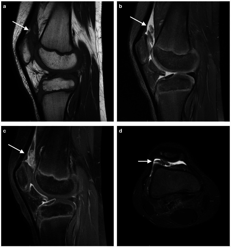
Pigmented villonodular synovitis (PVNS) is a relatively rare, benign lesion characterized by exuberant proliferation of the synovial tissue that most commonly affects the knee and hip joint. Magnetic resonance imaging (MRI) is the imaging modality of choice for the diagnosis of PVNS. The disease is confirmed histologically by examination of the synovial tissue removed. The mainstay of treatment is synovectomy, performed in an open, arthroscopic, or combined fashion. Although postoperative adjuvant external beam radiotherapy can improve the local recurrence rate, the course of the disease is not always uneventful. We present a rare case of a 10-year-old boy presented to our orthopaedic department with a four-month history of intermittent right knee pain and swelling. MRI revealed joint effusion and extensive nodular synovial proliferation suggestive of PVNS. An arthroscopic synovectomy was performed and histological examination confirmed the diagnosis. The postoperative course was uneventful. Clinical suspicion of PVNS is essential in children with chronic knee pain and swelling. Arthroscopic synovectomy is an effective and reliable treatment option.
色素沉着绒毛结节性滑膜炎(PVNS)是一种相对罕见的良性病变,其特征为滑膜组织的过度增生,最常累及膝关节和髋关节。磁共振成像(MRI)是诊断PVNS的首选影像学检查方法。通过对切除的滑膜组织进行组织学检查来确诊该病。主要治疗方法是滑膜切除术,可采用开放手术、关节镜手术或联合手术方式进行。尽管术后辅助外照射放疗可提高局部复发率,但疾病的病程并非总是顺利的。我们报告了一例罕见病例,一名10岁男孩因间歇性右膝疼痛和肿胀4个月就诊于我们的骨科。MRI显示有关节积液和广泛的结节状滑膜增生,提示PVNS。进行了关节镜下滑膜切除术,组织学检查确诊。术后病程顺利。对于有慢性膝关节疼痛和肿胀的儿童,临床怀疑PVNS至关重要。关节镜下滑膜切除术是一种有效且可靠的治疗选择。