Nishikawa Akihiro, Kakizawa Yukinari, Wada Naomichi, Yamamoto Yasunaga, Katsuki Masahito, Uchiyama Toshiya
Department of Neurosurgery, Suwa Red Cross Hospital, Suwa, Nagano, Japan.
World Neurosurg X. 2020 Dec 4;9:100096. doi: 10.1016/j.wnsx.2020.100096. eCollection 2021 Jan.
Time-of-flight magnetic resonance angiography (MRA) is limited by clip-induced artifacts after cerebral aneurysmal clipping. Recently, ultrashort echo time was shown to reduce metal artifacts. We assessed the pointwise encoding time reduction with radial acquisition (PETRA) sequence in subtraction-based MRA as an ultrashort echo time method during follow-up for clipping surgery.
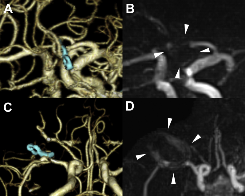
We retrospectively evaluated 114 branches of 63 aneurysms in 56 patients treated with titanium clips using MRA and 3-dimensional computed tomography angiography. The appearance using each method was compared, and the associations between visibility on PETRA-MRA, clip number and shape, and amount of hematoma were examined. Furthermore, the visibility of the aneurysm remnants and 2 clipping cases with cobalt-chromium-nickel-molybdenum clips were evaluated.
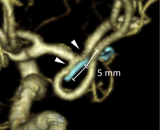
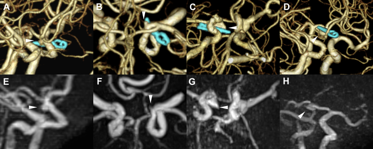
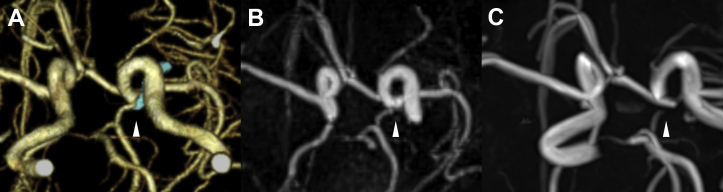
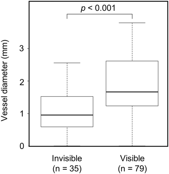
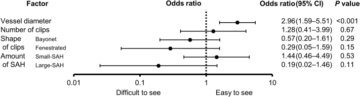
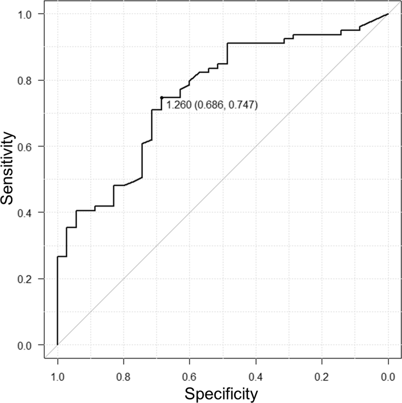
No branches were visible using time-of-flight-MRA, but 79 of 114 branches (69.3%) were visible on PETRA-MRA. PETRA-MRA was effective for follow-up imaging in 33 of 63 aneurysms (52.4%). The median vessel diameters were 1.67 mm (interquartile range, 1.24-2.62 mm) and 0.96 mm (interquartile range, 0.59-1.53 mm) in the visible and invisible groups, respectively. Only the vessel diameter correlated significantly ( < 0.001) with the visibility on PETRA-MRA. A receiver operating characteristic curve for the association between the vessel diameter and visibility on PETRA-MRA showed a cutoff value of 1.26 mm for vessel diameter. Cobalt-chromium-nickel-molybdenum clips produced a strong artifact, even on PETRA-MRA. All 4 residual aneurysms were visible on PETRA-MRA.
PETRA-MRA can be useful for follow-up aneurysm imaging when the diameter of vessels adjacent to the clip exceeds 1.26 mm. However, its usefulness is limited to titanium clips.
飞行时间磁共振血管造影(MRA)在脑动脉瘤夹闭术后受夹子引起的伪影限制。最近,超短回波时间已被证明可减少金属伪影。我们评估了基于减法的MRA中采用径向采集的逐点编码时间减少(PETRA)序列作为超短回波时间方法在夹闭手术随访中的应用。
我们回顾性评估了56例使用钛夹治疗的患者中63个动脉瘤的114个分支,采用MRA和三维计算机断层血管造影。比较了每种方法的表现,并研究了PETRA - MRA上的可见性、夹子数量和形状以及血肿量之间的关联。此外,评估了动脉瘤残余的可见性以及2例使用钴铬镍钼夹的夹闭病例。
飞行时间MRA未显示任何分支,但114个分支中的79个(69.3%)在PETRA - MRA上可见。PETRA - MRA对63个动脉瘤中的33个(52.4%)的随访成像有效。可见组和不可见组的血管直径中位数分别为1.67 mm(四分位间距,1.24 - 2.62 mm)和0.96 mm(四分位间距,0.59 - 1.53 mm)。只有血管直径与PETRA - MRA上的可见性显著相关(<0.001)。血管直径与PETRA - MRA上可见性之间关联的受试者工作特征曲线显示血管直径的截断值为1.26 mm。即使在PETRA - MRA上,钴铬镍钼夹也产生强烈伪影。所有4个残余动脉瘤在PETRA - MRA上均可见。
当夹附近血管直径超过1.26 mm时,PETRA - MRA可用于动脉瘤随访成像。然而,其用途仅限于钛夹。