Department of Biochemistry, Molecular Biology and Biophysics, University of Minnesota at Twin Cities, Minneapolis, MN 55455, USA.
Department of Pediatrics, University of Minnesota at Twin Cities, Minneapolis, MN 55455, USA.
Nutrients. 2021 Jan 8;13(1):179. doi: 10.3390/nu13010179.
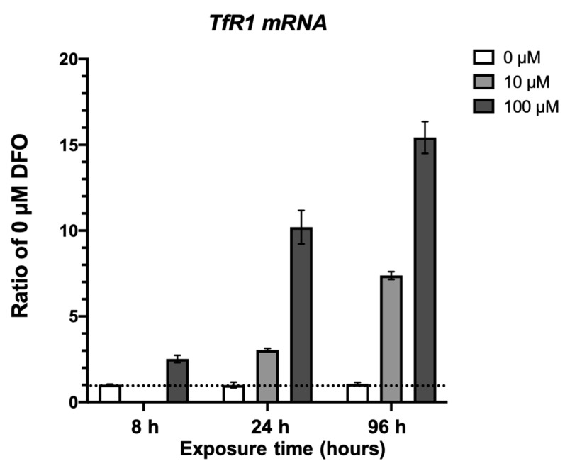
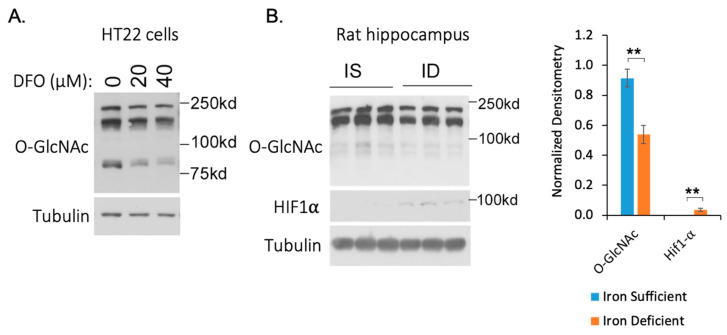
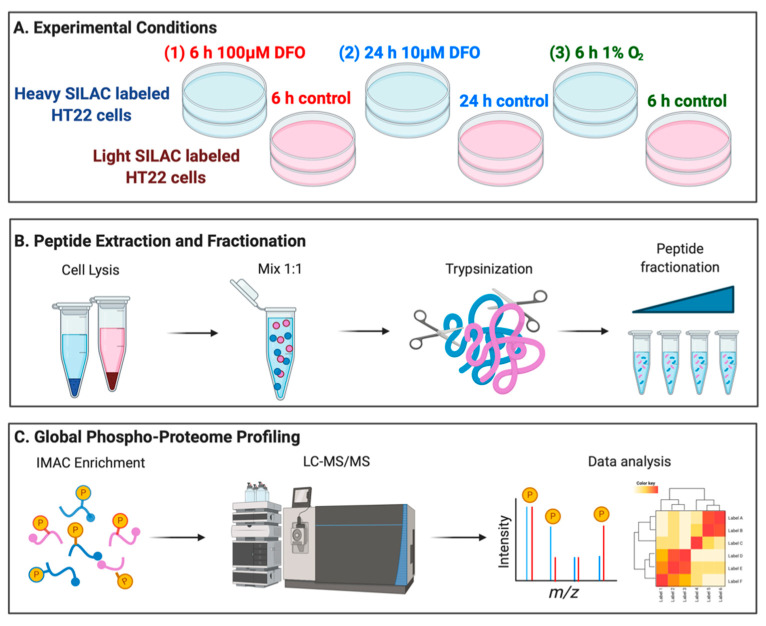
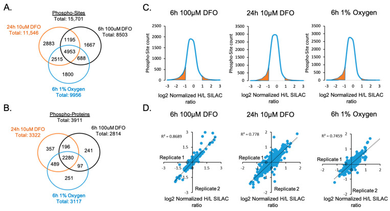
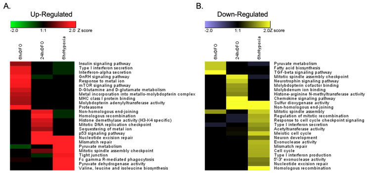
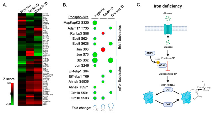
Micronutrient sensing is critical for cellular growth and differentiation. Deficiencies in essential nutrients such as iron strongly affect neuronal cell development and may lead to defects in neuronal function that cannot be remedied by subsequent iron supplementation. To understand the adaptive intracellular responses to iron deficiency in neuronal cells, we developed and utilized a Stable Isotopic Labeling of Amino acids in Cell culture (SILAC)-based quantitative phosphoproteomics workflow. Our integrated approach was designed to comprehensively elucidate the changes in phosphorylation signaling under both acute and chronic iron-deficient cell models. In addition, we analyzed the differential cellular responses between iron deficiency and hypoxia (oxygen-deprived) in neuronal cells. Our analysis identified nearly 16,000 phosphorylation sites in HT-22 cells, a hippocampal-derived neuronal cell line, more than ten percent of which showed at least 2-fold changes in response to either hypoxia or acute/chronic iron deficiency. Bioinformatic analysis revealed that iron deficiency altered key metabolic and epigenetic pathways including the phosphorylation of proteins involved in iron sequestration, glutamate metabolism, and histone methylation. In particular, iron deficiency increased glutamine-fructose-6-phosphate transaminase (GFPT1) phosphorylation, which is a key enzyme in the glucosamine biosynthesis pathway and a target of 5' AMP-activated protein kinase (AMPK), leading to reduced GFPT1 enzymatic activity and consequently lower global O-GlcNAc modification in neuronal cells. Taken together, our analysis of the phosphoproteome dynamics in response to iron and oxygen deprivation demonstrated an adaptive cellular response by mounting post-translational modifications that are critical for intracellular signaling and epigenetic programming in neuronal cells.
微量营养素感应对于细胞生长和分化至关重要。铁等必需营养素的缺乏会强烈影响神经元细胞的发育,并可能导致神经元功能的缺陷,而这些缺陷无法通过随后的铁补充来弥补。为了了解神经元细胞对铁缺乏的适应性细胞内反应,我们开发并利用了基于稳定同位素标记的氨基酸在细胞培养中的定量磷酸化蛋白质组学工作流程(SILAC)。我们的综合方法旨在全面阐明在急性和慢性铁缺乏细胞模型下磷酸化信号的变化。此外,我们分析了铁缺乏和缺氧(缺氧)对神经元细胞的差异细胞反应。我们的分析在 HT-22 细胞(海马源性神经元细胞系)中鉴定了近 16000 个磷酸化位点,其中超过 10%的磷酸化位点对缺氧或急性/慢性铁缺乏的反应至少有 2 倍的变化。生物信息学分析表明,铁缺乏改变了关键的代谢和表观遗传途径,包括铁螯合、谷氨酸代谢和组蛋白甲基化相关蛋白的磷酸化。特别是,铁缺乏增加了谷氨酰胺果糖-6-磷酸转氨酶(GFPT1)的磷酸化,GFPT1 是葡萄糖胺生物合成途径中的关键酶,也是 5'AMP 激活蛋白激酶(AMPK)的靶点,导致 GFPT1 酶活性降低,从而导致神经元细胞中全局 O-GlcNAc 修饰降低。总之,我们对铁和氧剥夺反应的磷酸蛋白质组动力学的分析表明,神经元细胞通过进行关键的细胞内信号和表观遗传编程的翻译后修饰来适应细胞反应。