• 文献检索
  • 文档翻译
  • 深度研究
  • 学术资讯
  • Suppr Zotero 插件Zotero 插件
  • 邀请有礼
  • 套餐&价格
  • 历史记录
应用&插件
Suppr Zotero 插件Zotero 插件浏览器插件Mac 客户端Windows 客户端微信小程序
定价
高级版会员购买积分包购买API积分包
服务
文献检索文档翻译深度研究API 文档MCP 服务
关于我们
关于 Suppr公司介绍联系我们用户协议隐私条款
关注我们

Suppr 超能文献

核心技术专利:CN118964589B侵权必究
粤ICP备2023148730 号-1Suppr @ 2026

文献检索

告别复杂PubMed语法,用中文像聊天一样搜索,搜遍4000万医学文献。AI智能推荐,让科研检索更轻松。

立即免费搜索

文件翻译

保留排版,准确专业,支持PDF/Word/PPT等文件格式,支持 12+语言互译。

免费翻译文档

深度研究

AI帮你快速写综述,25分钟生成高质量综述,智能提取关键信息,辅助科研写作。

立即免费体验

肝胆期肝脏病变呈高信号的谱系:基于临床背景的探讨方法

Spectrum of liver lesions hyperintense on hepatobiliary phase: an approach by clinical setting.

作者信息

Vernuccio Federica, Gagliano Domenico Salvatore, Cannella Roberto, Ba-Ssalamah Ahmed, Tang An, Brancatelli Giuseppe

机构信息

Department of Health Promotion, Mother and Child Care, Internal Medicine and Medical Specialties (PROMISE), University of Palermo, Via del Vespro, 129, 90127, Palermo, Italy.

University Paris Diderot, Sorbonne Paris Cité, Paris, France.

出版信息

Insights Imaging. 2021 Jan 12;12(1):8. doi: 10.1186/s13244-020-00928-w.

DOI:10.1186/s13244-020-00928-w
PMID:33432491
原文链接:https://pmc.ncbi.nlm.nih.gov/articles/PMC7801550/
Abstract

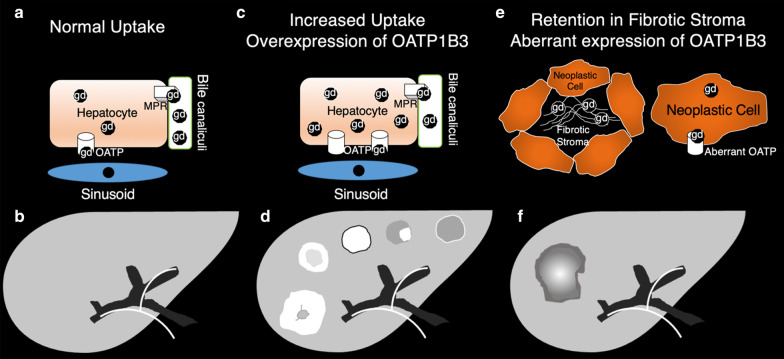
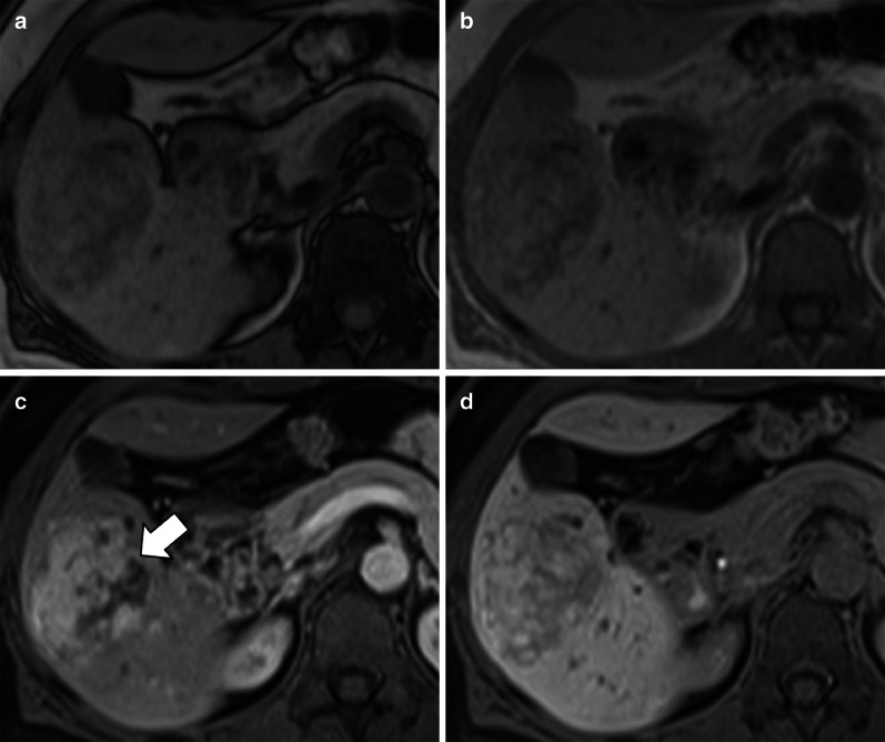
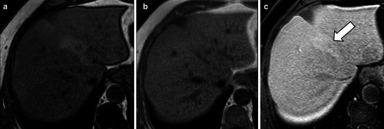
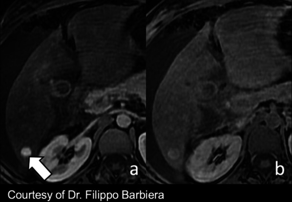
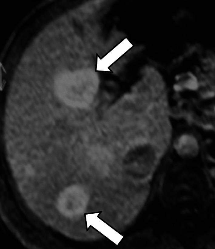
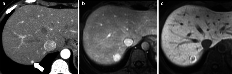
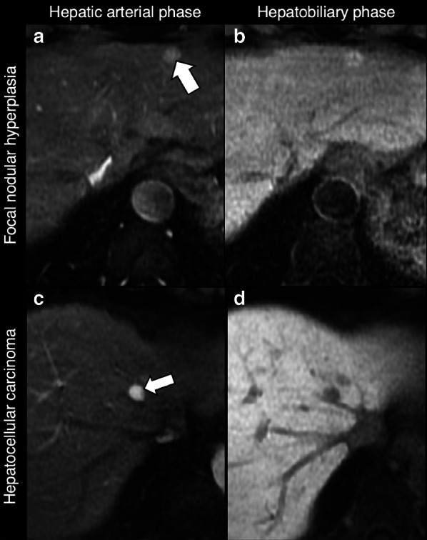
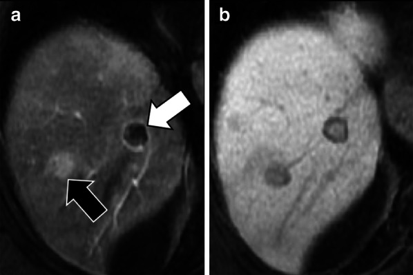
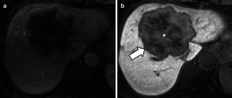
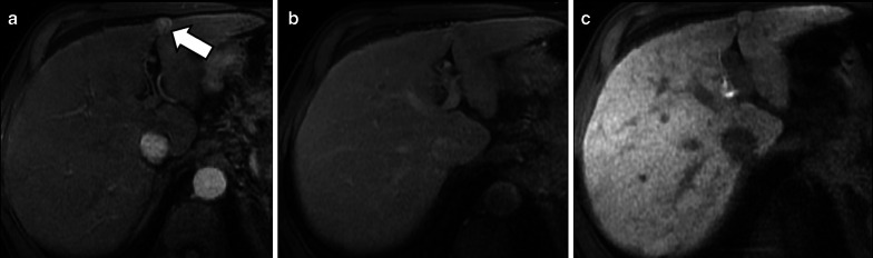
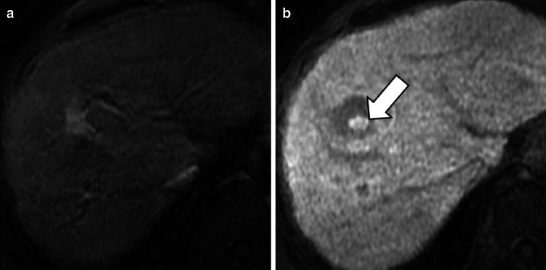
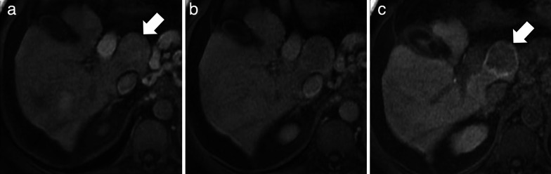
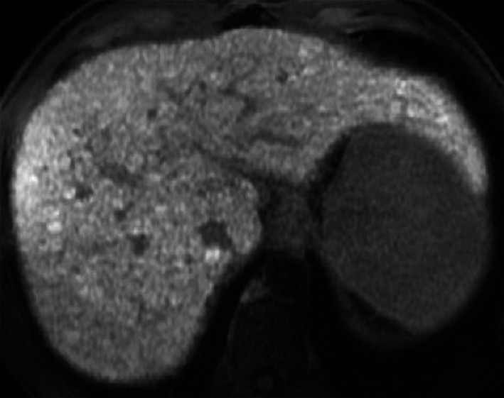
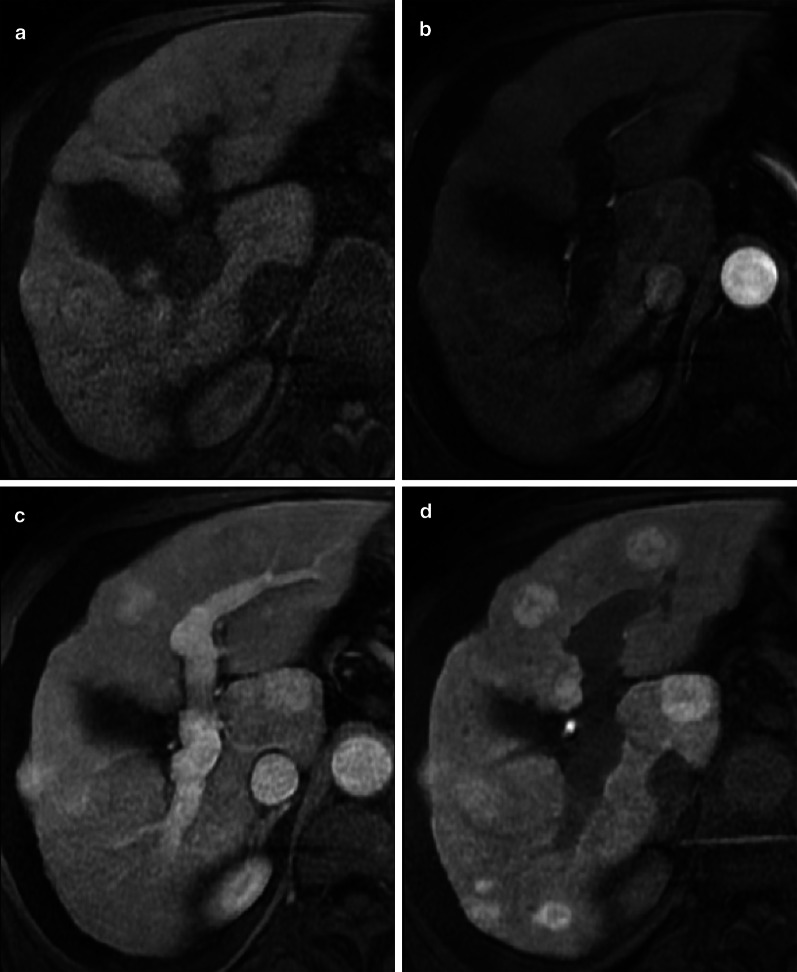
Hepatobiliary MRI contrast agents are increasingly being used for liver imaging. In clinical practice, most focal liver lesions do not uptake hepatobiliary contrast agents. Less commonly, hepatic lesions may show variable signal characteristics on hepatobiliary phase. This pictorial essay reviews a broad spectrum of benign and malignant focal hepatic observations that may show hyperintensity on hepatobiliary phase in various clinical settings. In non-cirrhotic patients, focal hepatic observations that show hyperintensity in the hepatobiliary phase are usually benign and typically include focal nodular hyperplasia. In patients with primary or secondary vascular disorders, focal nodular hyperplasia-like lesions arise as a local hyperplastic response to vascular alterations and tend to be iso- or hyperintense in the hepatobiliary phase. In oncologic patients, metastases and cholangiocarcinoma are hypointense lesions in the hepatobiliary phase; however, occasionally they may show a diffuse, central and inhomogeneous hepatobiliary paradoxical uptake with peripheral rim hypointensity. Post-chemotherapy focal nodular hyperplasia-like lesions may be tricky, and their typical hyperintense rim in the hepatobiliary phase is very helpful for the differential diagnosis with metastases. In cirrhotic patients, hepatocellular carcinoma may occasionally appear hyperintense on hepatobiliary phase.

摘要

肝胆磁共振成像造影剂越来越多地用于肝脏成像。在临床实践中,大多数肝脏局灶性病变不摄取肝胆造影剂。较少见的是,肝脏病变在肝胆期可能表现出不同的信号特征。这篇图文并茂的文章回顾了一系列广泛的良性和恶性肝脏局灶性表现,这些表现在各种临床情况下的肝胆期可能呈高信号。在非肝硬化患者中,在肝胆期呈高信号的肝脏局灶性表现通常是良性的,典型的包括局灶性结节性增生。在原发性或继发性血管疾病患者中,局灶性结节性增生样病变是对血管改变的局部增生反应,在肝胆期往往呈等信号或高信号。在肿瘤患者中,转移瘤和胆管癌在肝胆期是低信号病变;然而,偶尔它们可能表现出弥漫性、中央性和不均匀的肝胆矛盾性摄取,周边呈低信号。化疗后局灶性结节性增生样病变可能较难鉴别,其在肝胆期典型的高信号边缘对与转移瘤的鉴别诊断非常有帮助。在肝硬化患者中,肝细胞癌在肝胆期偶尔可能呈高信号。

https://cdn.ncbi.nlm.nih.gov/pmc/blobs/a660/7801550/e0106b7524b8/13244_2020_928_Fig17_HTML.jpg
https://cdn.ncbi.nlm.nih.gov/pmc/blobs/a660/7801550/fdadddb742bf/13244_2020_928_Fig1_HTML.jpg
https://cdn.ncbi.nlm.nih.gov/pmc/blobs/a660/7801550/7f660371ec62/13244_2020_928_Fig2_HTML.jpg
https://cdn.ncbi.nlm.nih.gov/pmc/blobs/a660/7801550/8791a7a27a0a/13244_2020_928_Fig3_HTML.jpg
https://cdn.ncbi.nlm.nih.gov/pmc/blobs/a660/7801550/47c64ce59e06/13244_2020_928_Fig4_HTML.jpg
https://cdn.ncbi.nlm.nih.gov/pmc/blobs/a660/7801550/983d3db88add/13244_2020_928_Fig5_HTML.jpg
https://cdn.ncbi.nlm.nih.gov/pmc/blobs/a660/7801550/68a6d428baba/13244_2020_928_Fig6_HTML.jpg
https://cdn.ncbi.nlm.nih.gov/pmc/blobs/a660/7801550/525c1ca0031c/13244_2020_928_Fig7_HTML.jpg
https://cdn.ncbi.nlm.nih.gov/pmc/blobs/a660/7801550/30c0192c1beb/13244_2020_928_Fig8_HTML.jpg
https://cdn.ncbi.nlm.nih.gov/pmc/blobs/a660/7801550/20ffb0ce7ce9/13244_2020_928_Fig9_HTML.jpg
https://cdn.ncbi.nlm.nih.gov/pmc/blobs/a660/7801550/2a0286f55eb0/13244_2020_928_Fig10_HTML.jpg
https://cdn.ncbi.nlm.nih.gov/pmc/blobs/a660/7801550/54dc4038677d/13244_2020_928_Fig11_HTML.jpg
https://cdn.ncbi.nlm.nih.gov/pmc/blobs/a660/7801550/132e31d46931/13244_2020_928_Fig12_HTML.jpg
https://cdn.ncbi.nlm.nih.gov/pmc/blobs/a660/7801550/adf7d118d15e/13244_2020_928_Fig13_HTML.jpg
https://cdn.ncbi.nlm.nih.gov/pmc/blobs/a660/7801550/bdd1ba4965f1/13244_2020_928_Fig14_HTML.jpg
https://cdn.ncbi.nlm.nih.gov/pmc/blobs/a660/7801550/53bce9ca7486/13244_2020_928_Fig15_HTML.jpg
https://cdn.ncbi.nlm.nih.gov/pmc/blobs/a660/7801550/6fe1633cbd25/13244_2020_928_Fig16_HTML.jpg
https://cdn.ncbi.nlm.nih.gov/pmc/blobs/a660/7801550/e0106b7524b8/13244_2020_928_Fig17_HTML.jpg

相似文献

1
Spectrum of liver lesions hyperintense on hepatobiliary phase: an approach by clinical setting.肝胆期肝脏病变呈高信号的谱系:基于临床背景的探讨方法
Insights Imaging. 2021 Jan 12;12(1):8. doi: 10.1186/s13244-020-00928-w.
2
Differentiation of hepatic hyperintense lesions seen on gadoxetic acid-enhanced hepatobiliary phase MRI.在钆塞酸增强肝胆期 MRI 上见到的肝脏高信号病变的鉴别诊断。
AJR Am J Roentgenol. 2011 Jul;197(1):W44-52. doi: 10.2214/AJR.10.5845.
3
Hypointense hepatic lesions depicted on gadobenate dimeglumine-enhanced three-hour delayed hepatobiliary-phase MR imaging: differentiation between benignancy and malignancy.钆贝葡胺增强三小时延迟肝胆期磁共振成像显示的肝脏低信号病变:良恶性鉴别
Korean J Radiol. 2009 May-Jun;10(3):294-302. doi: 10.3348/kjr.2009.10.3.294. Epub 2009 Apr 22.
4
Hyperintense lesions on gadoxetate disodium-enhanced hepatobiliary phase imaging.钆塞酸二钠增强肝胆期成像上的高信号病灶。
AJR Am J Roentgenol. 2012 Nov;199(5):W575-86. doi: 10.2214/AJR.11.8205.
5
Diagnosis of focal nodular hyperplasia with MRI: multicenter retrospective study comparing gadobenate dimeglumine to gadoxetate disodium.MRI 诊断局灶性结节性增生:对比钆贝葡胺与钆塞酸二钠的多中心回顾性研究。
AJR Am J Roentgenol. 2012 Jul;199(1):35-43. doi: 10.2214/AJR.11.7757.
6
Differentiation Between Hepatocellular Carcinoma Showing Hyperintensity on the Hepatobiliary Phase of Gadoxetic Acid-Enhanced MRI and Focal Nodular Hyperplasia by CT and MRI.CT 和 MRI 鉴别肝胆期增强 MRI 显示高信号的肝细胞癌与局灶性结节增生。
AJR Am J Roentgenol. 2018 Aug;211(2):347-357. doi: 10.2214/AJR.17.19341. Epub 2018 Apr 30.
7
Hepatobiliary MR contrast agents are useful to diagnose hepatocellular carcinoma in patients with Budd-Chiari syndrome.肝胆磁共振造影剂对布加综合征患者肝细胞癌的诊断很有用。
JHEP Rep. 2020 Mar 9;2(3):100097. doi: 10.1016/j.jhepr.2020.100097. eCollection 2020 Jun.
8
Gadoxetic acid-enhanced hepatobiliary phase MRI and high-b-value diffusion-weighted imaging to distinguish well-differentiated hepatocellular carcinomas from benign nodules in patients with chronic liver disease.钆塞酸增强肝胆期 MRI 和高 b 值扩散加权成像在慢性肝病患者中鉴别高分化肝细胞癌与良性结节
AJR Am J Roentgenol. 2011 Nov;197(5):W868-75. doi: 10.2214/AJR.10.6237.
9
MRI for characterization of benign hepatocellular tumors on hepatobiliary phase: the added value of in-phase imaging and lesion-to-liver visual signal intensity ratio.MRI 对肝胆期良恶性肝细胞肿瘤的特征分析:同相位成像和病变肝脏视觉信号强度比的附加价值。
Eur Radiol. 2019 Nov;29(11):5742-5751. doi: 10.1007/s00330-019-06210-y. Epub 2019 Apr 16.
10
Inflammatory hepatic adenomas: Characterization with hepatobiliary MRI contrast agents.炎症性肝腺瘤:使用肝胆磁共振成像造影剂的特征分析
Magn Reson Imaging. 2018 Apr;47:103-110. doi: 10.1016/j.mri.2017.12.006. Epub 2017 Dec 5.

引用本文的文献

1
Multiphase MRI radiomics model for predicting microvascular invasion in HCC: Development and clinical validation.用于预测肝癌微血管侵犯的多期MRI影像组学模型:开发与临床验证
ILIVER. 2025 Apr 26;4(2):100165. doi: 10.1016/j.iliver.2025.100165. eCollection 2025 Jun.
2
MRI major and ancillary features of LI-RADS for diagnosis of pediatric malignant liver tumors.用于诊断儿童恶性肝肿瘤的肝脏影像报告和数据系统(LI-RADS)的MRI主要及辅助特征。
Eur Radiol. 2025 Jun 25. doi: 10.1007/s00330-025-11775-y.
3
Intrahepatic diffuse periportal hyperintensity patterns on hepatobiliary phase of gadoxetate-enhanced MRI: a non-invasive imaging biomarker for clinical stratification of liver injury.

本文引用的文献

1
Hepatic enhancement in cirrhosis in the portal venous phase: what are the differences between gadoxetate disodium and gadobenate dimeglumine?肝硬化患者门静脉期肝实质增强:钆塞酸二钠与钆贝葡胺有何不同?
Abdom Radiol (NY). 2020 Aug;45(8):2409-2417. doi: 10.1007/s00261-020-02578-4.
2
Long-term Evolution of Hepatocellular Adenomas at MRI Follow-up.MRI 随访中肝细胞腺瘤的长期演变。
Radiology. 2020 May;295(2):361-372. doi: 10.1148/radiol.2020191790. Epub 2020 Mar 17.
3
Hepatocellular adenomas: is there additional value in using Gd-EOB-enhanced MRI for subtype differentiation?
钆塞酸增强MRI肝胆期肝内弥漫性门静脉周围高信号模式:一种用于肝损伤临床分层的非侵入性成像生物标志物。
Abdom Radiol (NY). 2025 May 13. doi: 10.1007/s00261-025-04985-x.
4
Intraindividual crossover comparison of gadobenate dimeglumine-enhanced and gadoxetate disodium-enhanced MRI for characterizing focal liver lesions.钆布醇与钆塞酸二钠增强MRI对肝脏局灶性病变特征的个体内交叉比较。
Eur Radiol Exp. 2025 Feb 18;9(1):23. doi: 10.1186/s41747-025-00551-8.
5
Imaging characteristics of hypervascular focal nodular hyperplasia-like lesions in patients with chronic alcoholic liver disease.慢性酒精性肝病患者中富血管性局灶性结节样增生性病变的影像学特征
World J Gastroenterol. 2025 Jan 14;31(2):98031. doi: 10.3748/wjg.v31.i2.98031.
6
MRI findings of newly present benign focal hepatic observations following chemotherapy: distinct features in early- and late-term follow-up.化疗后新出现的肝脏良性局灶性病变的MRI表现:早期和晚期随访中的不同特征
Eur Radiol. 2025 May;35(5):2783-2793. doi: 10.1007/s00330-024-11185-6. Epub 2024 Nov 12.
7
I thought it was a hemangioma! A pictorial essay about common and uncommon liver hemangiomas' mimickers.我原以为那是个血管瘤!一篇关于常见及罕见的肝脏血管瘤模仿者的图文并茂的文章。
Insights Imaging. 2024 Sep 19;15(1):228. doi: 10.1186/s13244-024-01745-1.
8
Discontinuous peripheral enhancement of focal liver lesions on CT and MRI: outside the box of typical cavernous hemangioma.CT和MRI上肝脏局灶性病变的不连续周边强化:超出典型海绵状血管瘤的范畴
Abdom Radiol (NY). 2025 Feb;50(2):693-709. doi: 10.1007/s00261-024-04522-2. Epub 2024 Aug 27.
9
Gd-EOB-DTPA enhanced MRI nomogram model to differentiate hepatocellular carcinoma and focal nodular hyperplasia both showing iso- or hyperintensity in the hepatobiliary phase.钆塞酸二钠增强 MRI 列线图模型鉴别肝胆期呈等或高信号的肝细胞肝癌和局灶性结节增生。
BMC Med Imaging. 2024 Aug 12;24(1):211. doi: 10.1186/s12880-024-01382-6.
10
Benign and malignant focal liver lesions displaying rim arterial phase hyperenhancement on CT and MRI.在CT和MRI上表现为边缘动脉期强化的肝脏局灶性良恶性病变。
Insights Imaging. 2024 Jul 18;15(1):178. doi: 10.1186/s13244-024-01756-y.
肝细胞腺瘤:钆塞酸二钠增强 MRI 在亚型鉴别方面是否具有附加价值?
Eur Radiol. 2020 Jun;30(6):3497-3506. doi: 10.1007/s00330-020-06726-8. Epub 2020 Feb 21.
4
Gadoxetic acid-enhanced MR imaging for hepatocellular carcinoma: molecular and genetic background.钆塞酸增强磁共振成像在肝细胞癌中的应用:分子和遗传背景。
Eur Radiol. 2020 Jun;30(6):3438-3447. doi: 10.1007/s00330-020-06687-y. Epub 2020 Feb 16.
5
Hyperintensity at fat spared area in steatotic liver on the hepatobiliary phase MRI.磁共振肝胆期成像中肝脂肪变区的脂肪信号保留区高信号。
Diagn Interv Radiol. 2019 Nov;25(6):416-420. doi: 10.5152/dir.2019.18535.
6
Hyperintense nodule-in-nodule on hepatobiliary phase arising within hypovascular hypointense nodule: Outcome and rate of hypervascular transformation.肝胆期呈低血供低信号结节内高信号结节:转归及高血供转化率。
Eur J Radiol. 2019 Nov;120:108689. doi: 10.1016/j.ejrad.2019.108689. Epub 2019 Sep 20.
7
CT and MR imaging findings of the livers in adults with Fontan palliation: an observational study.成人体外循环Fontan 术后肝脏的 CT 和 MR 成像表现:一项观察性研究。
Abdom Radiol (NY). 2020 Jan;45(1):188-202. doi: 10.1007/s00261-019-02119-8.
8
CT and MR imaging of chemotherapy-induced hepatopathy.化疗相关性肝损伤的 CT 和 MRI 成像。
Abdom Radiol (NY). 2019 Oct;44(10):3312-3324. doi: 10.1007/s00261-019-02193-y.
9
Diagnostic performance of gadoxetic acid-enhanced liver MRI versus multidetector CT in the assessment of colorectal liver metastases compared to hepatic resection.钆塞酸增强肝脏 MRI 与多排 CT 诊断结直肠癌肝转移与肝切除术后对比评估的性能比较。
BMC Gastroenterol. 2019 Jul 24;19(1):129. doi: 10.1186/s12876-019-1036-7.
10
Budd-Chiari Syndrome and hepatic regenerative nodules: Magnetic resonance findings with emphasis of hepatobiliary phase.布加综合征和肝再生结节:磁共振成像表现及肝胆期的重点。
Eur J Radiol. 2019 Aug;117:15-25. doi: 10.1016/j.ejrad.2019.05.015. Epub 2019 May 20.