Lea Dordi, Watson Martin, Skaland Ivar, Hagland Hanne R, Lillesand Melinda, Gudlaugsson Einar, Søreide Kjetil
Gastrointestinal Translational Research Unit, Molecular Laboratory, Hillevåg, Stavanger University Hospital, Stavanger, Norway.
Department of Clinical Medicine, University of Bergen, Bergen, Norway.
Cancer Immunol Immunother. 2021 Jul;70(7):2049-2057. doi: 10.1007/s00262-020-02834-y. Epub 2021 Jan 13.
In colon cancer, the location and density of tumor-infiltrating lymphocytes (TILs) can classify patients into low and high-risk groups for prognostication. While a commercially available 'Immunoscore' exists, the incurred expenses and copyrights may prevent universal use. The aim of this study was to develop a robust and objective quantification method of TILs in colon cancer.
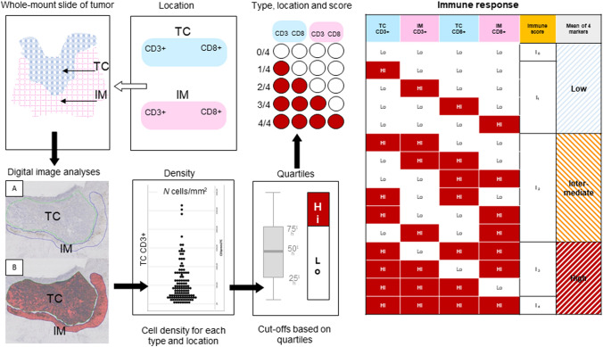
A consecutive, unselected series of specimens from patients with colon cancer were available for immunohistochemistry and assessment of TILs by automated digital pathology. CD3 + and CD8 + cells at the invasive margin and in tumor center were assessed on consecutive sections using automated digital pathology and image analysis software (Visiopharm). An algorithm template for whole slide assessment, generated cell counts per square millimeters (cells/mm), from which the immune score was calculated using distribution volumes. Furthermore, immune score was compared with clinical and histopathological characteristics to confirm its relevance.
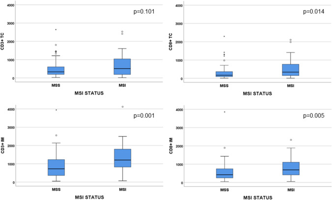
Based on the quantified TILs numbers by digital image analyses, patients were classified into low (n = 83, 69.7%), intermediate (n = 14, 11.8%) and high (n = 22, 18.5%) immune score groups. High immune score was associated with stage I-II tumors (p = 0.017) and a higher prevalence of microsatellite instable (MSI) tumors (p = 0.030). MSI tumors had a significantly higher numbers of CD3 + TILs in the invasive margin and CD8 + TILs in both tumor center and invasive margin, compared to microsatellite stable (MSS) tumors.
A digital template to quantify an easy-to-use immune score corresponds with clinicopathological features and MSI in colon cancer.
在结肠癌中,肿瘤浸润淋巴细胞(TILs)的位置和密度可将患者分为低风险和高风险预后组。虽然有一种商用的“免疫评分”,但其费用和版权可能会阻碍其广泛应用。本研究的目的是开发一种可靠且客观的结肠癌TILs定量方法。
连续选取未经筛选的结肠癌患者标本用于免疫组织化学,并通过自动数字病理学评估TILs。使用自动数字病理学和图像分析软件(Visiopharm)在连续切片上评估侵袭边缘和肿瘤中心的CD3+和CD8+细胞。生成了一个用于全切片评估的算法模板,计算每平方毫米的细胞计数(细胞/mm),并据此使用分布体积计算免疫评分。此外,将免疫评分与临床和组织病理学特征进行比较,以确认其相关性。
根据数字图像分析定量的TILs数量,患者被分为低免疫评分组(n = 83,69.7%)、中免疫评分组(n = 14,11.8%)和高免疫评分组(n = 22,18.5%)。高免疫评分与I-II期肿瘤相关(p = 0.017),且微卫星不稳定(MSI)肿瘤的患病率较高(p = 0.030)。与微卫星稳定(MSS)肿瘤相比,MSI肿瘤在侵袭边缘的CD3+ TILs数量以及在肿瘤中心和侵袭边缘的CD8+ TILs数量均显著更高。
一种用于量化易于使用的免疫评分的数字模板与结肠癌的临床病理特征和MSI相关。