Department of Ophthalmology and Visual Sciences, University of Wisconsin-Madison, Madison, WI, 53706, USA.
Department of Ophthalmology and Visual Sciences, University of Wisconsin-Madison, Madison, WI, 53706, USA; Department of Medical Microbiology and Immunology, University of Wisconsin-Madison, Madison, WI, 53706, USA; McPherson Eye Research Institute, University of Wisconsin-Madison, Madison, WI, 53706, USA.
Exp Eye Res. 2021 Mar;204:108436. doi: 10.1016/j.exer.2021.108436. Epub 2021 Jan 10.
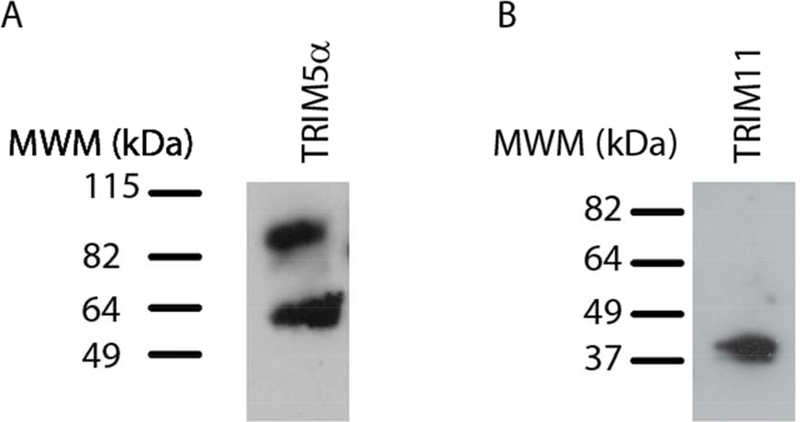
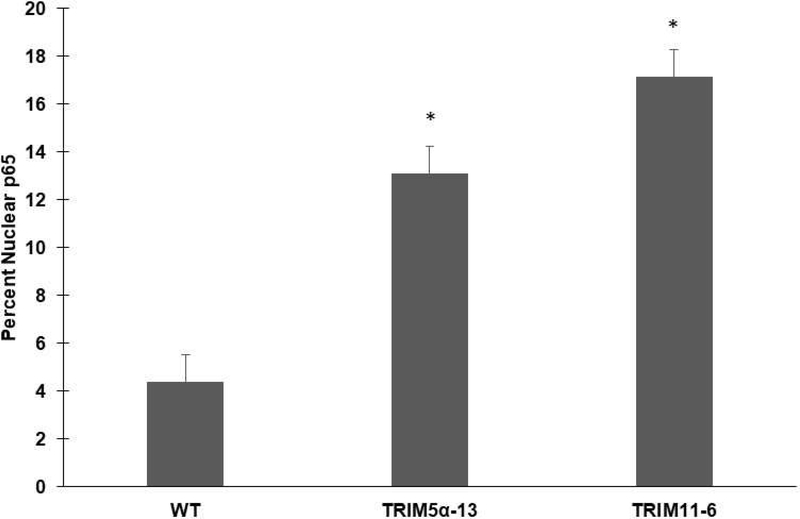
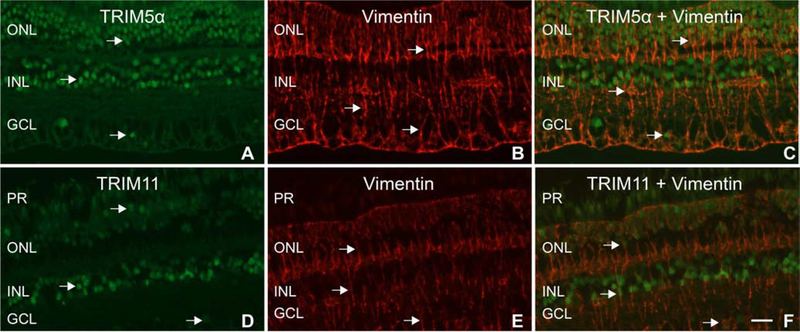
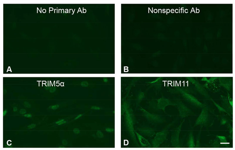
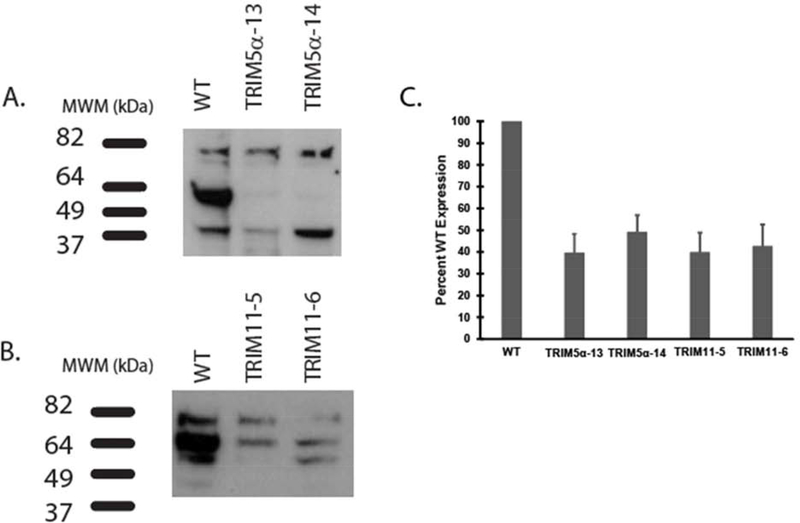
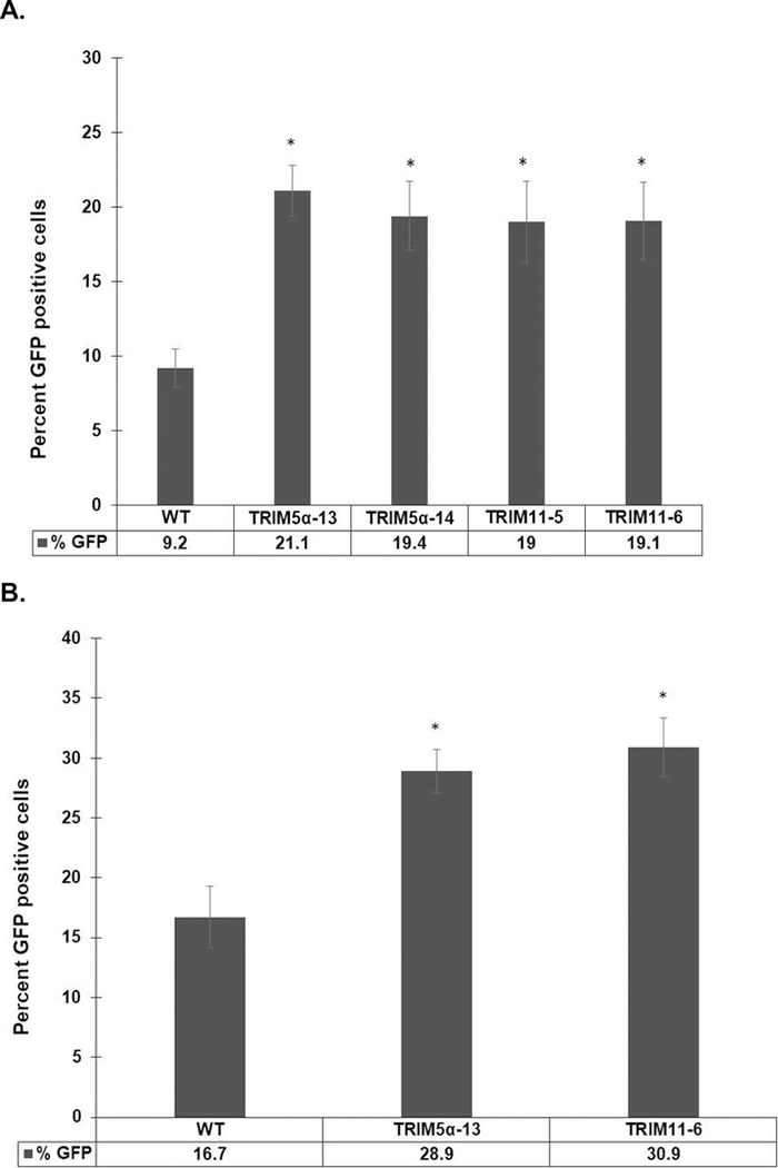
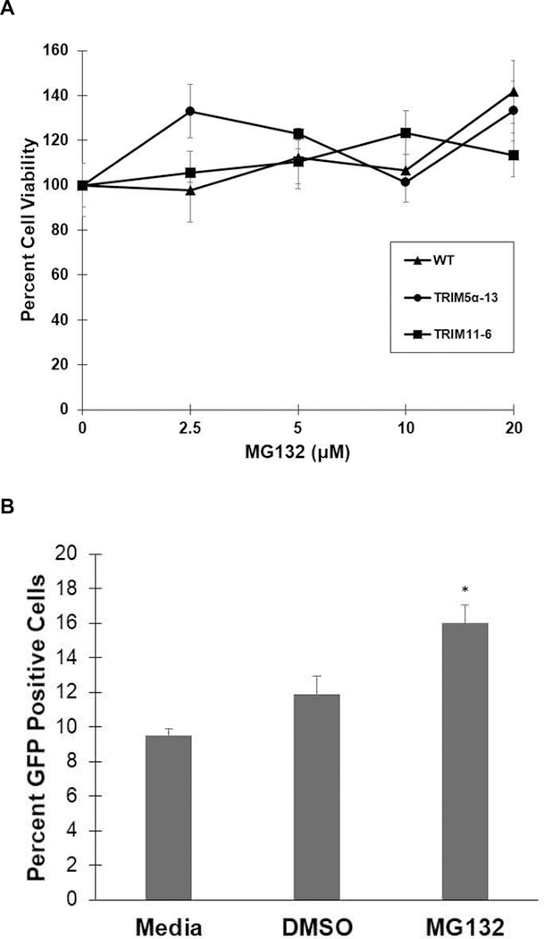
The goal of this study was to determine the expression and distribution of the host restriction factors (RFs) TRIM5α and TRIM11 in non-human primate (NHP) neural retina tissue and the human Muller cell line MIO-M1. In addition, experiments were performed to determine the effect of TRIM5α and TRIM11 knockdown on FIVGFP transduction of MIO-M1 cells with the goal of devising strategies to increase the efficiency of lentiviral (LV) gene delivery. Immunofluorescence (IF) studies indicated that TRIM5α and TRIM11 were localized predominantly in nuclei within the outer nuclear layer (ONL) and inner nuclear layer (INL) of NHP retina tissue. Double label IF indicated that TRIM5α and TRIM11 were localized to some of the retinal Muller cell nuclei. MIO-M1 cells expressed TRIM5α predominantly in the nucleus and TRIM11 primarily in the cytosol. FIVGFP transduction efficiency was significantly increased, at 4 and 7 days post transduction, in TRIM5α and TRIM11 knockdown clones (KD) compared to WT MIO-M1 cells. In addition, pretreatment with the proteasome inhibitor MG132 increased the transduction efficiency of FIVGFP in WT MIO-M1 cells. The nuclear translocation of NF-κB (p65), at 72 h post FIVGFP transduction, was enhanced in TRIM5α and TRIM11 KD clones. The expression of TRIM5α and TRIM11 in macaque neural retina tissue and MIO-M1 cells indicate the presence of these RFs in NHP retina and human Muller cells. Our data indicate that even partial knockdown of TRIM5α or TRIM11, or a short proteasome inhibitor pretreatment, can increase the transduction efficiency of a LV vector.
本研究旨在确定宿主限制因子(RFs)TRIM5α 和 TRIM11 在非人类灵长类动物(NHP)神经视网膜组织和人 Muller 细胞系 MIO-M1 中的表达和分布。此外,还进行了实验以确定 TRIM5α 和 TRIM11 敲低对 MIO-M1 细胞中 FIVGFP 转导的影响,目的是设计提高慢病毒(LV)基因传递效率的策略。免疫荧光(IF)研究表明,TRIM5α 和 TRIM11 主要位于 NHP 视网膜组织的外核层(ONL)和内核层(INL)的核内。双标记 IF 表明,TRIM5α 和 TRIM11 定位于一些视网膜 Muller 细胞核内。MIO-M1 细胞主要在核内表达 TRIM5α,主要在细胞质中表达 TRIM11。与 WT MIO-M1 细胞相比,在 TRIM5α 和 TRIM11 敲低克隆(KD)中转导 4 天和 7 天后,FIVGFP 的转导效率显着提高。此外,用蛋白酶体抑制剂 MG132 预处理可增加 WT MIO-M1 细胞中 FIVGFP 的转导效率。在 FIVGFP 转导后 72 小时,TRIM5α 和 TRIM11 KD 克隆中 NF-κB(p65)的核易位增强。Macaque 神经视网膜组织和 MIO-M1 细胞中 TRIM5α 和 TRIM11 的表达表明这些 RF 存在于 NHP 视网膜和人 Muller 细胞中。我们的数据表明,即使是 TRIM5α 或 TRIM11 的部分敲低,或短暂的蛋白酶体抑制剂预处理,也可以提高 LV 载体的转导效率。