• 文献检索
  • 文档翻译
  • 深度研究
  • 学术资讯
  • Suppr Zotero 插件Zotero 插件
  • 邀请有礼
  • 套餐&价格
  • 历史记录
应用&插件
Suppr Zotero 插件Zotero 插件浏览器插件Mac 客户端Windows 客户端微信小程序
定价
高级版会员购买积分包购买API积分包
服务
文献检索文档翻译深度研究API 文档MCP 服务
关于我们
关于 Suppr公司介绍联系我们用户协议隐私条款
关注我们

Suppr 超能文献

核心技术专利:CN118964589B侵权必究
粤ICP备2023148730 号-1Suppr @ 2026

文献检索

告别复杂PubMed语法,用中文像聊天一样搜索,搜遍4000万医学文献。AI智能推荐,让科研检索更轻松。

立即免费搜索

文件翻译

保留排版,准确专业,支持PDF/Word/PPT等文件格式,支持 12+语言互译。

免费翻译文档

深度研究

AI帮你快速写综述,25分钟生成高质量综述,智能提取关键信息,辅助科研写作。

立即免费体验

圆锥角膜交联术后12个月进展期圆锥角膜患者的角膜地形图结果

Outcomes of Corneal Topography among Progressive Keratoconus Patients 12 months following Corneal Collagen Cross-Linking.

作者信息

AlQahtani Bader S, Alshahrani Saeed, Khayyat Waleed W, Abdalla-Elsayed Maram E A, Altalhi Abdullah A, Saifaldein Amjad A, Taha Mohammed A

机构信息

College of Medicine, King Saud Bin Abdulaziz University for Health Sciences, Riyadh, Saudi Arabia.

King Abdullah International Medical Research Center, Riyadh, Saudi Arabia.

出版信息

Clin Ophthalmol. 2021 Jan 7;15:49-55. doi: 10.2147/OPTH.S284981. eCollection 2021.

DOI:10.2147/OPTH.S284981
PMID:33442228
原文链接:https://pmc.ncbi.nlm.nih.gov/articles/PMC7800689/
Abstract

PURPOSE

This study aimed to assess the overall and specific topographic changes among patients who underwent corneal collagen cross-linking (CXL) due to progressive keratoconus.

METHODS

This retrospective case series study was conducted at a single-arm hospital in King Abdulaziz Medical City, Riyadh. All progressive keratoconus patients who underwent CXL between January 2017 and December 2018 were included consecutively. The epi-off crosslinking technique (Dresden protocol) was applied in all patients. The topographic values were measured preoperatively and 12 months postoperatively. Patients with a history of a previous corneal procedure, corneal trauma, or any corneal scarring were excluded.

RESULTS

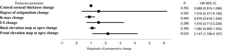
Among our population (29 eyes of 24 patients), 58.6% of eyes were for male patients, and the mean age of the population was 27.76 ± 4.21 years. Based on the topography results, the mean values of corneal thickness at central 3 mm decreased from 473.45 ± 38 µm to 465.72 ± 41.78 µm following CXL (Z = -1.93, 95% confidence interval [CI] = 0.048-0.057, p= 0.053). Clinically significant astigmatism measurements were present in 28 (96.6%) eyes before CXL compared to 26 (89.7%) eyes after CXL. The mean values of astigmatism among the patients were 3.37 ± 2.25 diopters before and 3.67 ± 2.61 diopters after CXL (Z = -1696, 95% confidence interval [CI] = 0.085-0.096, p = 0.09). After CXL, the mean values of the front elevation at the apex changed from 33.90 ± 20.13 µm to 36.10 ± 21.09 µm (Z = -2.792, 95% [CI] = 0.003-0.006, p = 0.005). The mean values of the back elevation at the apex changed from 68.4 ± 35.66 µm to 69.90 ± 35.89 µm (Z = -0.934, 95% CI = 0.343-0.366, p = 0.35).

CONCLUSION

The topographic corneal parameters improved significantly in the patients with corneal ectasia after CXL. These results revealed the safety and efficacy of CXL in stabilizing keratoconus progression among Saudi patients at 1 year of follow-up.

摘要

目的

本研究旨在评估因进行性圆锥角膜而接受角膜胶原交联(CXL)的患者的整体和特定地形变化。

方法

本回顾性病例系列研究在利雅得阿卜杜勒阿齐兹国王医疗城的一家单臂医院进行。连续纳入2017年1月至2018年12月期间接受CXL的所有进行性圆锥角膜患者。所有患者均采用上皮移除交联技术(德累斯顿方案)。术前和术后12个月测量地形值。排除有既往角膜手术、角膜外伤或任何角膜瘢痕病史的患者。

结果

在我们的研究人群(24例患者的29只眼)中,58.6%的眼为男性患者,人群平均年龄为27.76±4.21岁。根据地形结果,CXL后中央3mm处角膜厚度的平均值从473.45±38μm降至465.72±41.78μm(Z=-1.93,95%置信区间[CI]=0.048-0.057,p=0.053)。CXL前28只眼(96.6%)存在临床显著散光测量值,而CXL后为26只眼(89.7%)。患者散光平均值术前为3.37±2.25屈光度,术后为3.67±2.61屈光度(Z=-1696,95%置信区间[CI]=0.085-0.096,p=0.09)。CXL后,顶点处前表面高度的平均值从33.90±20.13μm变为36.10±21.09μm(Z=-2.792,95%[CI]=0.003-0.006,p=0.005)。顶点处后表面高度的平均值从68.4±35.66μm变为69.90±35.89μm(Z=-0.934,95%CI=0.343-0.366,p=0.35)。

结论

CXL后角膜扩张患者的地形角膜参数有显著改善。这些结果揭示了CXL在沙特患者随访1年时稳定圆锥角膜进展方面的安全性和有效性。

https://cdn.ncbi.nlm.nih.gov/pmc/blobs/89c8/7800689/8c866a911e96/OPTH-15-49-g0001.jpg

相似文献

1
Outcomes of Corneal Topography among Progressive Keratoconus Patients 12 months following Corneal Collagen Cross-Linking.圆锥角膜交联术后12个月进展期圆锥角膜患者的角膜地形图结果
Clin Ophthalmol. 2021 Jan 7;15:49-55. doi: 10.2147/OPTH.S284981. eCollection 2021.
2
Outcomes of Epi-On Collagen Cross-Linkage Procedure Assessed in Progressive Keratoconus Patients.在进展性圆锥角膜患者中评估的上皮瓣下胶原交联手术的结果。
Cureus. 2022 Oct 25;14(10):e30664. doi: 10.7759/cureus.30664. eCollection 2022 Oct.
3
[Predictive factors for visual outcome after corneal collagen crosslinking treatment in progressive keratoconus: One-year refractive and topographic results].[圆锥角膜交联治疗后视力预后的预测因素:一年屈光和地形图结果]
J Fr Ophtalmol. 2015 Sep;38(7):595-606. doi: 10.1016/j.jfo.2014.11.017. Epub 2015 May 18.
4
Comparison of transepithelial corneal collagen crosslinking with epithelium-off crosslinking in progressive keratoconus.进行性圆锥角膜中经上皮角膜胶原交联与去上皮交联的比较。
J Fr Ophtalmol. 2014 May;37(5):371-6. doi: 10.1016/j.jfo.2013.11.012. Epub 2014 Mar 26.
5
Corneal Collagen Cross-Linking for Keratoconus in Pediatric Patients-Long-Term Results.儿童圆锥角膜的角膜胶原交联术——长期结果
Cornea. 2017 Feb;36(2):138-143. doi: 10.1097/ICO.0000000000001102.
6
Iontophoretic collagen cross-linking versus epithelium-off collagen cross-linking for early stage of progressive keratoconus - 3 years follow-up study.离子导入胶原交联与上皮去除胶原交联治疗早期进行性圆锥角膜的3年随访研究
Acta Ophthalmol. 2017 Nov;95(7):e649-e655. doi: 10.1111/aos.13538.
7
Corneal collagen cross-linking with riboflavin and ultraviolet a irradiation for keratoconus: long-term results.角膜胶原交联联合核黄素和紫外线 A 照射治疗圆锥角膜:长期疗效。
Ophthalmology. 2013 Aug;120(8):1515-20. doi: 10.1016/j.ophtha.2013.01.012. Epub 2013 Apr 12.
8
Risk factors for keratoconus progression after treatment by accelerated cross-linking (A-CXL): A prospective 24-month study.加速交联(A-CXL)治疗后圆锥角膜进展的危险因素:一项为期24个月的前瞻性研究。
J Fr Ophtalmol. 2021 Jun;44(6):863-872. doi: 10.1016/j.jfo.2020.08.040. Epub 2021 May 28.
9
Epithelium-Off vs. transepithelial corneal collagen crosslinking in progressive keratoconus: 3 years of follow-up.上皮下型与经上皮型角膜胶原交联术治疗进展性圆锥角膜:3 年随访结果。
J Optom. 2021 Apr-Jun;14(2):189-198. doi: 10.1016/j.optom.2020.07.005. Epub 2020 Sep 1.
10
Long-term visual, refractive, tomographic and aberrometric outcomes of corneal collagen crosslinking (CXL) with or without hypoosmolar riboflavin solution in the treatment of progressive keratoconus patients with thin corneas.角膜胶原交联术(CXL)联合或不联合低渗核黄素溶液治疗薄角膜进展性圆锥角膜患者的长期视力、屈光、断层和像差结果。
Graefes Arch Clin Exp Ophthalmol. 2022 Apr;260(4):1225-1235. doi: 10.1007/s00417-021-05314-w. Epub 2021 Nov 27.

引用本文的文献

1
Digital transformation of care for keratoconus patients: ML modeling structural outcomes of corneal collagen cross-linking.圆锥角膜患者护理的数字化转型:角膜胶原交联的机器学习建模结构结果
Front Med (Lausanne). 2025 Jun 4;12:1462653. doi: 10.3389/fmed.2025.1462653. eCollection 2025.
2
Quantifying choroidal and retinal thicknesses variations via optical coherence tomography in different stages of pediatric keratoconus.通过光学相干断层扫描定量分析不同阶段儿童圆锥角膜的脉络膜和视网膜厚度变化。
BMC Ophthalmol. 2024 Nov 7;24(1):488. doi: 10.1186/s12886-024-03753-7.
3
Outcomes of Corneal Topographical Kmax readings 3 months after Corneal Cross Linkage in Keratoconus patients.

本文引用的文献

1
Pachymetry and Elevation Back Map Changes in Keratoconus Patients After Crosslinking Procedure.交联术后圆锥角膜患者角膜厚度和高度背侧地图变化。
Med Arch. 2020 Apr;74(2):105-108. doi: 10.5455/medarh.2020.74.105-108.
2
Prevalence of Subclinical Keratoconus Among Pediatric Egyptian Population with Astigmatism.埃及散光儿童人群中亚临床圆锥角膜的患病率
Clin Ophthalmol. 2020 Mar 23;14:905-913. doi: 10.2147/OPTH.S245492. eCollection 2020.
3
Prevalence of keratoconus in paediatric patients in Riyadh, Saudi Arabia.沙特阿拉伯利雅得儿科患者圆锥角膜的患病率。
圆锥角膜患者角膜交联术后3个月角膜地形图Kmax读数的结果
J Community Hosp Intern Med Perspect. 2022 Sep 9;12(5):43-47. doi: 10.55729/2000-9666.1111. eCollection 2022.
4
The 25th Anniversary of Laser Vision Correction in the United States.美国激光视力矫正25周年。
Clin Ophthalmol. 2021 Mar 17;15:1163-1172. doi: 10.2147/OPTH.S299752. eCollection 2021.
Br J Ophthalmol. 2018 Oct;102(10):1436-1441. doi: 10.1136/bjophthalmol-2017-311391. Epub 2018 Jan 3.
4
Corneal Changes After Collagen Crosslinking for Keratoconus Using Dual Scheimpflug Imaging.使用双Scheimpflug成像技术对圆锥角膜进行胶原交联后的角膜变化
J Ophthalmic Vis Res. 2015 Oct-Dec;10(4):358-63. doi: 10.4103/2008-322X.176894.
5
Different Topographic Response Between Mild to Moderate and Advanced Keratoconus After Accelerated Collagen Cross-linking.加速角膜交联术后轻度至中度和重度圆锥角膜的不同地形图反应
Cornea. 2015 Aug;34(8):922-7. doi: 10.1097/ICO.0000000000000483.
6
Changes in corneal topography and biomechanical properties after collagen cross linking for keratoconus: 1-year results.圆锥角膜胶原交联术后角膜地形图和生物力学特性的变化:1年结果
Middle East Afr J Ophthalmol. 2015 Apr-Jun;22(2):212-9. doi: 10.4103/0974-9233.151877.
7
A review of collagen cross-linking in cornea and sclera.角膜和巩膜中胶原交联的综述。
J Ophthalmol. 2015;2015:289467. doi: 10.1155/2015/289467. Epub 2015 Apr 2.
8
Pentacam topographic changes after collagen cross-linking in patients with keratoconus.圆锥角膜患者胶原交联术后的Pentacam地形图变化
Adv Biomed Res. 2015 Feb 23;4:62. doi: 10.4103/2277-9175.151886. eCollection 2015.
9
Corneal collagen cross-linking in keratoconus: a systematic review and meta-analysis.圆锥角膜的角膜胶原交联:一项系统评价和荟萃分析。
Sci Rep. 2014 Jul 10;4:5652. doi: 10.1038/srep05652.
10
Corneal topography patterns in the Tehran eye study: warning about the high prevalence of patterns with a skewed radial axis.德黑兰眼科研究中的角膜地形图模式:关于径向轴偏斜模式高患病率的警示
Middle East Afr J Ophthalmol. 2014 Jan-Mar;21(1):72-6. doi: 10.4103/0974-9233.124107.