• 文献检索
  • 文档翻译
  • 深度研究
  • 学术资讯
  • Suppr Zotero 插件Zotero 插件
  • 邀请有礼
  • 套餐&价格
  • 历史记录
应用&插件
Suppr Zotero 插件Zotero 插件浏览器插件Mac 客户端Windows 客户端微信小程序
定价
高级版会员购买积分包购买API积分包
服务
文献检索文档翻译深度研究API 文档MCP 服务
关于我们
关于 Suppr公司介绍联系我们用户协议隐私条款
关注我们

Suppr 超能文献

核心技术专利:CN118964589B侵权必究
粤ICP备2023148730 号-1Suppr @ 2026

文献检索

告别复杂PubMed语法,用中文像聊天一样搜索,搜遍4000万医学文献。AI智能推荐,让科研检索更轻松。

立即免费搜索

文件翻译

保留排版,准确专业,支持PDF/Word/PPT等文件格式,支持 12+语言互译。

免费翻译文档

深度研究

AI帮你快速写综述,25分钟生成高质量综述,智能提取关键信息,辅助科研写作。

立即免费体验

相似文献

1
Enhanced Proliferation of Ly6C Monocytes/Macrophages Contributes to Chronic Inflammation in Skin Wounds of Diabetic Mice.Ly6C 单核细胞/巨噬细胞的增殖增强导致糖尿病小鼠皮肤伤口慢性炎症。
J Immunol. 2021 Feb 1;206(3):621-630. doi: 10.4049/jimmunol.2000935. Epub 2020 Dec 21.
2
Proliferation of Ly6C+ monocytes/macrophages contributes to their accumulation in mouse skin wounds.Ly6C+ 单核细胞/巨噬细胞的增殖有助于其在小鼠皮肤伤口中的积累。
J Leukoc Biol. 2020 Apr;107(4):551-560. doi: 10.1002/JLB.3HI1119-389RRRR. Epub 2019 Nov 28.
3
Reduced apoptosis of monocytes and macrophages is associated with their persistence in wounds of diabetic mice.单核细胞和巨噬细胞凋亡减少与它们在糖尿病小鼠伤口中的持续存在有关。
Cytokine. 2021 Jun;142:155516. doi: 10.1016/j.cyto.2021.155516. Epub 2021 Mar 31.
4
Ly6C Blood Monocyte/Macrophage Drive Chronic Inflammation and Impair Wound Healing in Diabetes Mellitus.Ly6C 血液单核细胞/巨噬细胞驱动糖尿病慢性炎症并损害伤口愈合。
Arterioscler Thromb Vasc Biol. 2018 May;38(5):1102-1114. doi: 10.1161/ATVBAHA.118.310703. Epub 2018 Mar 1.
5
Spatio-temporal profile, phenotypic diversity, and fate of recruited monocytes into the post-ischemic brain.募集单核细胞进入缺血后大脑的时空特征、表型多样性及转归
J Neuroinflammation. 2016 Nov 4;13(1):285. doi: 10.1186/s12974-016-0750-0.
6
Ly6CLo Monocyte/Macrophages are Essential for Thrombus Resolution in a Murine Model of Venous Thrombosis.Ly6CLo 单核细胞/巨噬细胞对于静脉血栓形成的小鼠模型中的血栓溶解是必不可少的。
Thromb Haemost. 2020 Feb;120(2):289-299. doi: 10.1055/s-0039-3400959. Epub 2019 Dec 30.
7
CCR2 Deficiency Impairs Ly6C and Ly6C Monocyte Responses in Infection.CCR2缺陷损害感染过程中Ly6C和Ly6C单核细胞反应。
Front Immunol. 2021 Jul 5;12:670219. doi: 10.3389/fimmu.2021.670219. eCollection 2021.
8
Macrophage dynamics are regulated by local macrophage proliferation and monocyte recruitment in injured pancreas.巨噬细胞动力学受损伤胰腺中局部巨噬细胞增殖和单核细胞募集的调节。
Eur J Immunol. 2015 May;45(5):1482-93. doi: 10.1002/eji.201445013. Epub 2015 Mar 19.
9
Origin of Monocytes/Macrophages Contributing to Chronic Inflammation in Chagas Disease: SIRT1 Inhibition of FAK-NFκB-Dependent Proliferation and Proinflammatory Activation of Macrophages.导致恰加斯病慢性炎症的单核细胞/巨噬细胞的起源:SIRT1 抑制 FAk-NFκB 依赖性增殖和巨噬细胞的促炎激活。
Cells. 2019 Dec 28;9(1):80. doi: 10.3390/cells9010080.
10
Resident and pro-inflammatory macrophages in the colon represent alternative context-dependent fates of the same Ly6Chi monocyte precursors.结肠中的驻留和促炎巨噬细胞代表了相同 Ly6Chi 单核细胞前体在不同环境条件下的两种替代命运。
Mucosal Immunol. 2013 May;6(3):498-510. doi: 10.1038/mi.2012.89. Epub 2012 Sep 19.

引用本文的文献

1
The spatial and temporal activation of macrophages during fibrosis.纤维化过程中巨噬细胞的时空激活
Nat Rev Immunol. 2025 Jun 4. doi: 10.1038/s41577-025-01186-x.
2
Increased ROS and Persistent Pro-Inflammatory Responses in a Diabetic Wound Healing Model (db/db): Implications for Delayed Wound Healing.糖尿病伤口愈合模型(db/db)中活性氧增加及持续的促炎反应:对伤口愈合延迟的影响
Int J Mol Sci. 2025 May 20;26(10):4884. doi: 10.3390/ijms26104884.
3
The Role of Omics Techniques in Diabetic Wound Healing: Recent Insights into the Application of Single-Cell RNA Sequencing, Bulk RNA Sequencing, Spatial Transcriptomics, and Proteomics.组学技术在糖尿病伤口愈合中的作用:单细胞RNA测序、全转录组RNA测序、空间转录组学和蛋白质组学应用的最新见解
Adv Ther. 2025 May 17. doi: 10.1007/s12325-025-03212-9.
4
Anti?atherosclerotic effect of aged garlic extract: Mode of action and therapeutic benefits (Review).陈年大蒜提取物的抗动脉粥样硬化作用:作用方式及治疗益处(综述)
Exp Ther Med. 2025 Mar 26;29(5):104. doi: 10.3892/etm.2025.12854. eCollection 2025 May.
5
CDK1-loaded extracellular vesicles promote cell cycle to reverse impaired wound healing in diabetic obese mice.携带细胞周期蛋白依赖性激酶1的细胞外囊泡可促进细胞周期,逆转糖尿病肥胖小鼠受损的伤口愈合。
Mol Ther. 2025 Mar 5;33(3):1118-1133. doi: 10.1016/j.ymthe.2025.01.039. Epub 2025 Jan 25.
6
Elucidating the dual roles of apoptosis and necroptosis in diabetic wound healing: implications for therapeutic intervention.阐明细胞凋亡和坏死性凋亡在糖尿病伤口愈合中的双重作用:对治疗干预的启示。
Burns Trauma. 2025 Jan 22;13:tkae061. doi: 10.1093/burnst/tkae061. eCollection 2025.
7
Characterizing Fibroblast Heterogeneity in Diabetic Wounds Through Single-Cell RNA-Sequencing.通过单细胞RNA测序表征糖尿病伤口中的成纤维细胞异质性
Biomedicines. 2024 Nov 7;12(11):2538. doi: 10.3390/biomedicines12112538.
8
Fibroblast-Mediated Macrophage Recruitment Supports Acute Wound Healing.成纤维细胞介导的巨噬细胞募集促进急性伤口愈合。
J Invest Dermatol. 2024 Nov 22. doi: 10.1016/j.jid.2024.10.609.
9
The role of adipose tissue-derived stromal cells, macrophages and bioscaffolds in cutaneous wound repair.脂肪组织来源的基质细胞、巨噬细胞和生物支架在皮肤伤口修复中的作用。
Biol Direct. 2024 Sep 29;19(1):85. doi: 10.1186/s13062-024-00534-6.
10
Histone modifications and their roles in macrophage-mediated inflammation: a new target for diabetic wound healing.组蛋白修饰及其在巨噬细胞介导的炎症中的作用:糖尿病创面愈合的新靶点。
Front Immunol. 2024 Aug 20;15:1450440. doi: 10.3389/fimmu.2024.1450440. eCollection 2024.

本文引用的文献

1
Proliferation of Ly6C+ monocytes/macrophages contributes to their accumulation in mouse skin wounds.Ly6C+ 单核细胞/巨噬细胞的增殖有助于其在小鼠皮肤伤口中的积累。
J Leukoc Biol. 2020 Apr;107(4):551-560. doi: 10.1002/JLB.3HI1119-389RRRR. Epub 2019 Nov 28.
2
Trends in incidence of total or type 2 diabetes: systematic review.糖尿病发病率的趋势:系统综述。
BMJ. 2019 Sep 11;366:l5003. doi: 10.1136/bmj.l5003.
3
Calmodulin-dependent signalling pathways are activated and mediate the acute inflammatory response of injured skeletal muscle.钙调蛋白依赖性信号通路被激活并介导受损骨骼肌的急性炎症反应。
J Physiol. 2019 Nov;597(21):5161-5177. doi: 10.1113/JP278478. Epub 2019 Oct 10.
4
Cx3CR1 Expression Identifies Distinct Macrophage Populations That Contribute Differentially to Inflammation and Repair.Cx3CR1表达可识别对炎症和修复有不同贡献的不同巨噬细胞群体。
Immunohorizons. 2019 Jul 1;3(7):262-273. doi: 10.4049/immunohorizons.1900038.
5
Diabetes induces myeloid bias in bone marrow progenitors associated with enhanced wound macrophage accumulation and impaired healing.糖尿病导致骨髓祖细胞中的髓样偏倚,与伤口巨噬细胞积累增加和愈合受损有关。
J Pathol. 2019 Dec;249(4):435-446. doi: 10.1002/path.5330. Epub 2019 Aug 28.
6
CCL2-Mediated Reversal of Impaired Skin Wound Healing in Diabetic Mice by Normalization of Neovascularization and Collagen Accumulation.CCL2 介导体外诱导糖尿病小鼠皮肤伤口愈合的实验研究
J Invest Dermatol. 2019 Dec;139(12):2517-2527.e5. doi: 10.1016/j.jid.2019.05.022. Epub 2019 Jun 24.
7
Recruitment of hepatic macrophages from monocytes is independent of IL-4Rα but is associated with ablation of resident macrophages in schistosomiasis.从单核细胞中招募肝巨噬细胞不依赖于 IL-4Rα,但与血吸虫病中固有巨噬细胞的清除有关。
Eur J Immunol. 2019 Jul;49(7):1067-1081. doi: 10.1002/eji.201847796. Epub 2019 Apr 24.
8
Astrocytes-derived VEGF exacerbates the microvascular damage of late delayed RBI.星形胶质细胞衍生的 VEGF 加剧了迟发性 RBI 的微血管损伤。
Neuroscience. 2019 Jun 1;408:14-21. doi: 10.1016/j.neuroscience.2019.03.039. Epub 2019 Mar 22.
9
Current health and economic burden of chronic diabetic osteomyelitis.慢性糖尿病性骨髓炎的当前健康和经济负担。
Expert Rev Pharmacoecon Outcomes Res. 2019 Jun;19(3):279-286. doi: 10.1080/14737167.2019.1567337. Epub 2019 Jan 21.
10
Loss of MD1 exacerbates myocardial ischemia/reperfusion injury and susceptibility to ventricular arrhythmia.MD1 的缺失会加重心肌缺血/再灌注损伤和易发性室性心律失常。
Eur J Pharmacol. 2019 Feb 5;844:79-86. doi: 10.1016/j.ejphar.2018.11.025. Epub 2018 Nov 17.

Ly6C 单核细胞/巨噬细胞的增殖增强导致糖尿病小鼠皮肤伤口慢性炎症。

Enhanced Proliferation of Ly6C Monocytes/Macrophages Contributes to Chronic Inflammation in Skin Wounds of Diabetic Mice.

机构信息

Center for Wound Healing and Tissue Regeneration, Department of Kinesiology and Nutrition, University of Illinois at Chicago, Chicago, IL 60612; and.

Research Informatics Core, University of Illinois at Chicago, Chicago, IL 60612.

出版信息

J Immunol. 2021 Feb 1;206(3):621-630. doi: 10.4049/jimmunol.2000935. Epub 2020 Dec 21.

DOI:10.4049/jimmunol.2000935
PMID:33443065
原文链接:https://pmc.ncbi.nlm.nih.gov/articles/PMC7927918/
Abstract

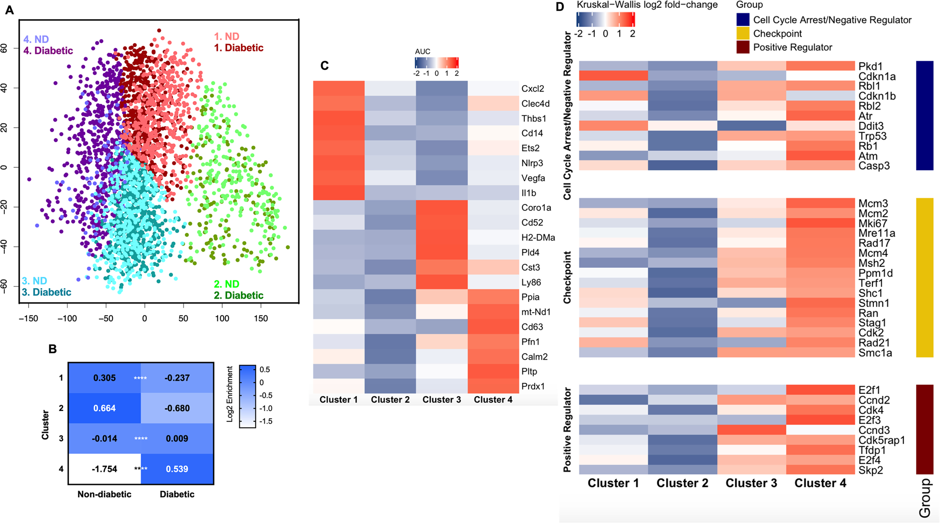
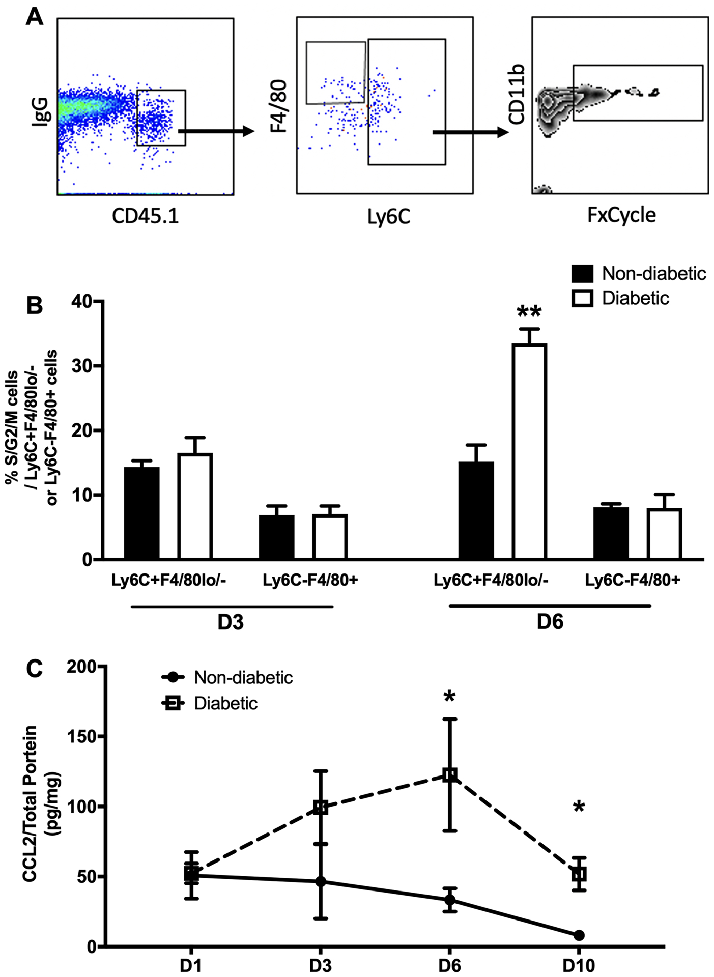
Diabetic wounds are characterized by persistent accumulation of proinflammatory monocytes (Mo)/macrophages (MΦ) and impaired healing. However, the mechanisms underlying the persistent accumulation of Mo/MΦ remain poorly understood. In this study, we report that Ly6CF4/80 Mo/MΦ proliferate at higher rates in wounds of diabetic mice compared with nondiabetic mice, leading to greater accumulation of these cells. Unbiased single cell RNA sequencing analysis of combined nondiabetic and diabetic wound Mo/MΦ revealed a cluster, populated primarily by cells from diabetic wounds, for which genes associated with the cell cycle were enriched. In a screen of potential regulators, CCL2 levels were increased in wounds of diabetic mice, and subsequent experiments showed that local CCL2 treatment increased Ly6CF4/80 Mo/MΦ proliferation. Importantly, adoptive transfer of mixtures of CCR2 and CCR2 Ly6C Mo indicated that CCL2/CCR2 signaling is required for their proliferation in the wound environment. Together, these data demonstrate a novel role for the CCL2/CCR2 signaling pathway in promoting skin Mo/MΦ proliferation, contributing to persistent accumulation of Mo/MΦ and impaired healing in diabetic mice.

摘要

糖尿病伤口的特征是促炎单核细胞(Mo)/巨噬细胞(MΦ)持续积累和愈合受损。然而,Mo/MΦ持续积累的机制仍知之甚少。在这项研究中,我们报告称,与非糖尿病小鼠相比,糖尿病小鼠伤口中的 Ly6CF4/80 Mo/MΦ 以更高的速度增殖,导致这些细胞的积累更多。对非糖尿病和糖尿病伤口 Mo/MΦ 的组合进行无偏单细胞 RNA 测序分析显示,一个主要由来自糖尿病伤口的细胞组成的簇,其与细胞周期相关的基因富集。在对潜在调节剂的筛选中,糖尿病小鼠伤口中的 CCL2 水平升高,随后的实验表明局部 CCL2 治疗可增加 Ly6CF4/80 Mo/MΦ 的增殖。重要的是,CCR2 和 CCR2 Ly6C Mo 的混合物的过继转移表明,CCL2/CCR2 信号通路是其在伤口环境中增殖所必需的。总之,这些数据表明 CCL2/CCR2 信号通路在促进皮肤 Mo/MΦ 增殖方面具有新的作用,导致糖尿病小鼠 Mo/MΦ 的持续积累和愈合受损。

https://cdn.ncbi.nlm.nih.gov/pmc/blobs/c4e6/7927918/2f535b58ff3c/nihms-1650343-f0005.jpg
https://cdn.ncbi.nlm.nih.gov/pmc/blobs/c4e6/7927918/155bc8ee0ccc/nihms-1650343-f0001.jpg
https://cdn.ncbi.nlm.nih.gov/pmc/blobs/c4e6/7927918/f8714ad8d0b3/nihms-1650343-f0002.jpg
https://cdn.ncbi.nlm.nih.gov/pmc/blobs/c4e6/7927918/a97db61d9715/nihms-1650343-f0003.jpg
https://cdn.ncbi.nlm.nih.gov/pmc/blobs/c4e6/7927918/a719c57118e8/nihms-1650343-f0004.jpg
https://cdn.ncbi.nlm.nih.gov/pmc/blobs/c4e6/7927918/2f535b58ff3c/nihms-1650343-f0005.jpg