Bristol Medical School, Faculty of Health Sciences, University of Bristol, Bristol, BS1 3NY, UK.
Neuroscience and Mental Health Research Institute, Cardiff University, Cardiff, CF24 4HQ, UK.
Brain Stimul. 2021 Mar-Apr;14(2):217-225. doi: 10.1016/j.brs.2021.01.003. Epub 2021 Jan 12.
Transcranial ultrasound stimulation can acutely modulate brain activity, but the lasting effects on neurons are unknown.
To assess the excitability profile of neurons in the hours following transient ultrasound stimulation.
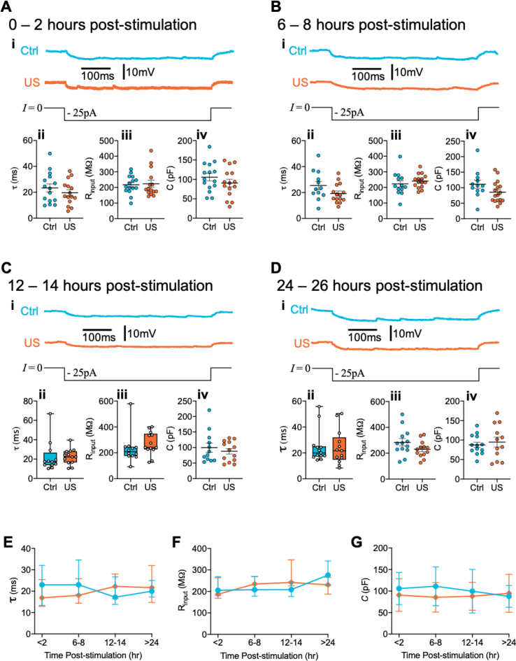
Primary rat cortical neurons were stimulated with a 40 s, 200 kHz pulsed ultrasound stimulation or sham-stimulation. Intrinsic firing properties were investigated through whole-cell patch-clamp recording by evoking action potentials in response to somatic current injection. Recordings were taken at set timepoints following ultrasound stimulation: 0-2 h, 6-8 h, 12-14 h and 24-26 h. Transmission electron microscopy was used to assess synaptic ultrastructure at the same timepoints.
In the 0-2 h window, neurons stimulated with ultrasound displayed an increase in the mean frequency of evoked action potentials of 32% above control cell levels (p = 0.023). After 4-6 h this increase was measured as 44% (p = 0.0043). By 12-14 h this effect was eliminated and remained absent 24-26 h post-stimulation. These changes to action potential firing occurred in conjunction with statistically significant differences between control and ultrasound-stimulated neurons in action potential half-width, depolarisation rate, and repolarisation rate, that were similarly eliminated by 24 h following stimulation. These effects occurred in the absence of alterations to intrinsic membrane properties or synaptic ultrastructure.
We report that stimulating neurons with 40 s of ultrasound enhances their excitability for up to 8 h in conjunction with modifications to action potential kinetics. This occurs in the absence of major ultrastructural change or modification of intrinsic membrane properties. These results can inform the application of transcranial ultrasound in experimental and therapeutic settings.
经颅超声刺激可以急性调节大脑活动,但对神经元的持续影响尚不清楚。
评估短暂超声刺激后数小时神经元的兴奋性特征。
使用 40 秒、200 kHz 脉冲超声刺激或假刺激刺激原代大鼠皮质神经元。通过在体电流注入引发动作电位来研究全细胞膜片钳记录中的固有放电特性。在超声刺激后的设定时间点进行记录:0-2 小时、6-8 小时、12-14 小时和 24-26 小时。在相同的时间点使用透射电子显微镜评估突触超微结构。
在 0-2 小时的窗口中,与对照细胞水平相比,经超声刺激的神经元显示出 32%的诱发动作电位平均频率增加(p=0.023)。4-6 小时后,这一增加被测量为 44%(p=0.0043)。到 12-14 小时,这种效应消失,刺激后 24-26 小时仍未出现。这些动作电位放电的变化伴随着控制和超声刺激神经元在动作电位半宽度、去极化率和复极化率方面的统计学显著差异,这些差异在刺激后 24 小时也被消除。这些效应发生在不改变内在膜特性或突触超微结构的情况下。
我们报告,用 40 秒的超声刺激神经元可在 8 小时内增强其兴奋性,同时改变动作电位动力学。这是在没有主要超微结构改变或内在膜特性改变的情况下发生的。这些结果可以为经颅超声在实验和治疗环境中的应用提供信息。