• 文献检索
  • 文档翻译
  • 深度研究
  • 学术资讯
  • Suppr Zotero 插件Zotero 插件
  • 邀请有礼
  • 套餐&价格
  • 历史记录
应用&插件
Suppr Zotero 插件Zotero 插件浏览器插件Mac 客户端Windows 客户端微信小程序
定价
高级版会员购买积分包购买API积分包
服务
文献检索文档翻译深度研究API 文档MCP 服务
关于我们
关于 Suppr公司介绍联系我们用户协议隐私条款
关注我们

Suppr 超能文献

核心技术专利:CN118964589B侵权必究
粤ICP备2023148730 号-1Suppr @ 2026

文献检索

告别复杂PubMed语法,用中文像聊天一样搜索,搜遍4000万医学文献。AI智能推荐,让科研检索更轻松。

立即免费搜索

文件翻译

保留排版,准确专业,支持PDF/Word/PPT等文件格式,支持 12+语言互译。

免费翻译文档

深度研究

AI帮你快速写综述,25分钟生成高质量综述,智能提取关键信息,辅助科研写作。

立即免费体验

肾母细胞瘤1(WT1):隆突性皮肤纤维肉瘤的一种新型免疫标志物——对114例外观平淡的真皮/皮下组织间质性梭形细胞病变的免疫组织化学研究

Wilms' Tumor 1 (WT1): A Novel Immunomarker of Dermatofibrosarcoma Protuberans-An Immunohistochemical Study on a Series of 114 Cases of Bland-Looking Mesenchymal Spindle Cell Lesions of the Dermis/Subcutaneous Tissues.

作者信息

Piombino Eliana, Broggi Giuseppe, Barbareschi Mattia, Castorina Sergio, Parenti Rosalba, Bartoloni Giovanni, Salvatorelli Lucia, Magro Gaetano

机构信息

Department of Medical and Surgical Sciences and Advanced Technologies, G.F. Ingrassia, Azienda Ospedaliero-Universitaria "Policlinico Vittorio Emanuele", Anatomic Pathology, School of Medicine, University of Catania, 95123 Catania, Italy.

Pathology Unit, Department of Clinical Services, Santa Chiara Hospital, 38122 Trento, Italy.

出版信息

Cancers (Basel). 2021 Jan 12;13(2):252. doi: 10.3390/cancers13020252.

DOI:10.3390/cancers13020252
PMID:33445443
原文链接:https://pmc.ncbi.nlm.nih.gov/articles/PMC7826654/
Abstract

PURPOSE

to investigate the immunohistochemical expression and distribution of Wilms' tumor 1 (WT1) (transcription factor produced by the tumor suppressor gene of the same name) in a series of 114 cases of bland-looking mesenchymal spindle cell lesions of the dermis/subcutaneous tissues to establish whether this immunomarker is differentially expressed in dermatofibrosarcoma protuberans (DFSP) versus its potential morphological mimickers.

METHODS

This retrospective multi-centric immunohistochemical study included 57 DFSP cases, 15 dermatofibromas, 5 deep fibrous histiocytomas, 8 neurofibromas, 5 spindle cell lipomas, 8 dermal scars, 6 nodular fasciitis, 5 cutaneous leiomyomas and 5 solitary fibrous tumors. Among the 57 DFSP cases, 11 were recurrent lesions; 2 non-recurrent cases exhibited an additional "" overgrowth and 1 recurrent and 2 primary tumors contained a minority of "" components.

RESULTS

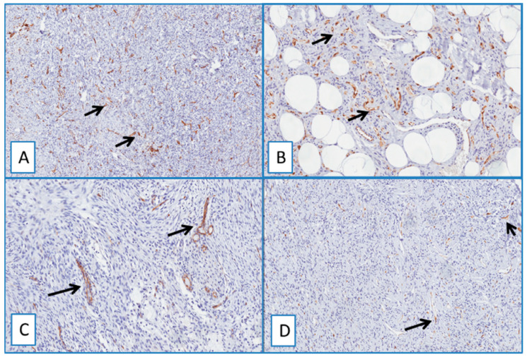
Most DFSP (95% of cases) exhibited cytoplasmic staining for WT1; 11/11 residual/recurrent tumors showed diffuse and strong WT1 cytoplasmic immunoreactivity; apart from neurofibromas, WT1 expression was lacking in all the other cases studied.

CONCLUSIONS

The cytoplasmic expression of WT1 may be exploitable as a complementary diagnostic immunomarker to CD34 in confirming the diagnosis of DFSP and to better evaluate the residual/recurrent tumor component.

摘要

目的

研究威尔姆斯瘤1(WT1)(由同名肿瘤抑制基因产生的转录因子)在114例外观平淡的真皮/皮下组织间质性梭形细胞病变中的免疫组化表达及分布情况,以确定该免疫标志物在隆突性皮肤纤维肉瘤(DFSP)及其潜在形态学模仿者中是否存在差异表达。

方法

这项回顾性多中心免疫组化研究包括57例DFSP病例、15例皮肤纤维瘤、5例深部纤维组织细胞瘤、8例神经纤维瘤、5例梭形细胞脂肪瘤、8例皮肤瘢痕、6例结节性筋膜炎、5例皮肤平滑肌瘤和5例孤立性纤维瘤。在57例DFSP病例中,11例为复发病变;2例非复发病例表现出额外的“过度生长”,1例复发病例和2例原发性肿瘤含有少数“”成分。

结果

大多数DFSP(95%的病例)表现出WT1的细胞质染色;11/11例残留/复发性肿瘤显示WT1细胞质免疫反应弥漫且强烈;除神经纤维瘤外,所有其他研究病例均缺乏WT1表达。

结论

WT1的细胞质表达可作为CD34的补充诊断免疫标志物,用于确诊DFSP并更好地评估残留/复发性肿瘤成分。

https://cdn.ncbi.nlm.nih.gov/pmc/blobs/c485/7826654/791e97a10f96/cancers-13-00252-g005.jpg
https://cdn.ncbi.nlm.nih.gov/pmc/blobs/c485/7826654/a08afe672f6d/cancers-13-00252-g001.jpg
https://cdn.ncbi.nlm.nih.gov/pmc/blobs/c485/7826654/ae60dbfa6cbb/cancers-13-00252-g002.jpg
https://cdn.ncbi.nlm.nih.gov/pmc/blobs/c485/7826654/81cfec567d0f/cancers-13-00252-g003.jpg
https://cdn.ncbi.nlm.nih.gov/pmc/blobs/c485/7826654/3d411cdb43fe/cancers-13-00252-g004.jpg
https://cdn.ncbi.nlm.nih.gov/pmc/blobs/c485/7826654/791e97a10f96/cancers-13-00252-g005.jpg

相似文献

1
Wilms' Tumor 1 (WT1): A Novel Immunomarker of Dermatofibrosarcoma Protuberans-An Immunohistochemical Study on a Series of 114 Cases of Bland-Looking Mesenchymal Spindle Cell Lesions of the Dermis/Subcutaneous Tissues.肾母细胞瘤1(WT1):隆突性皮肤纤维肉瘤的一种新型免疫标志物——对114例外观平淡的真皮/皮下组织间质性梭形细胞病变的免疫组织化学研究
Cancers (Basel). 2021 Jan 12;13(2):252. doi: 10.3390/cancers13020252.
2
Cytoplasmic expression of Wilms tumor transcription factor-1 (WT1): a useful immunomarker for young-type fibromatoses and infantile fibrosarcoma.肾母细胞瘤转录因子-1(WT1)的细胞质表达:一种用于幼年型纤维瘤病和婴儿纤维肉瘤的有用免疫标志物。
Acta Histochem. 2014 Sep;116(7):1134-40. doi: 10.1016/j.acthis.2014.05.010. Epub 2014 Jul 5.
3
Fibrosarcomatous ("high-grade") dermatofibrosarcoma protuberans: clinicopathologic and immunohistochemical study of a series of 41 cases with emphasis on prognostic significance.纤维肉瘤样(“高级别”)隆突性皮肤纤维肉瘤:41例系列病例的临床病理及免疫组化研究,重点关注预后意义。
Am J Surg Pathol. 1998 May;22(5):576-87. doi: 10.1097/00000478-199805000-00009.
4
Dermatofibrosarcoma Protuberans: An Immunomarker Study of 57 Cases That Included Putative Mesenchymal Stem Cell Markers.隆突性皮肤纤维肉瘤:一项包含假定间充质干细胞标志物的57例病例的免疫标志物研究。
Appl Immunohistochem Mol Morphol. 2017 Sep;25(8):586-591. doi: 10.1097/PAI.0000000000000348.
5
Subcutaneous dermatofibrosarcoma protuberans, a rare subtype with predilection for the head: A retrospective series of 18 cases.皮下隆突性皮肤纤维肉瘤,一种罕见的好发于头面部的亚型:回顾性系列研究 18 例。
J Am Acad Dermatol. 2017 Sep;77(3):503-511.e1. doi: 10.1016/j.jaad.2017.02.046. Epub 2017 Apr 15.
6
Differential expression of HMGA1 and HMGA2 in dermatofibroma and dermatofibrosarcoma protuberans: potential diagnostic applications, and comparison with histologic findings, CD34, and factor XIIIa immunoreactivity.HMGA1和HMGA2在皮肤纤维瘤和隆突性皮肤纤维肉瘤中的差异表达:潜在的诊断应用,以及与组织学结果、CD34和凝血因子XIIIa免疫反应性的比较
Am J Dermatopathol. 2004 Aug;26(4):267-72. doi: 10.1097/00000372-200408000-00001.
7
Dermatofibrosarcoma protuberans, giant cell fibroblastoma, and hybrid lesions in children: clinicopathologic comparative analysis of 28 cases with molecular data--a study from the French Federation of Cancer Centers Sarcoma Group.儿童隆突性皮肤纤维肉瘤、巨细胞成纤维细胞瘤及混合性病变:28例临床病理比较分析及分子数据——来自法国癌症中心肉瘤研究组的一项研究
Am J Surg Pathol. 2003 Jan;27(1):27-39. doi: 10.1097/00000478-200301000-00004.
8
[Dermatofibrosarcoma Darier and Ferrand. Immunohistochemistry study of 38 cases. Research on a specific marker].[达里埃和费朗的皮肤纤维肉瘤。38例免疫组织化学研究。特定标志物的研究]
Ann Pathol. 1997 Oct;17(5):324-8.
9
Fibrosarcomatous dermatofibrosarcoma protuberans with giant rosettes.伴有巨大玫瑰花结的纤维肉瘤样隆突性皮肤纤维肉瘤
Am J Dermatopathol. 2001 Feb;23(1):41-5. doi: 10.1097/00000372-200102000-00007.
10
Giant cell fibroblastoma: a report of three cases with histologic and immunohistochemical evidence of a relationship to dermatofibrosarcoma protuberans.巨细胞成纤维细胞瘤:三例报告,其组织学和免疫组化证据表明与隆突性皮肤纤维肉瘤相关。
Arch Pathol Lab Med. 1996 Nov;120(11):1052-5.

引用本文的文献

1
Pleomorphic dermal sarcoma of the scalp with intracranial space involvement: management of a rare entity. Illustrative case.头皮多形性皮肤肉瘤伴颅内间隙受累:罕见病例的处理。病例说明
J Neurosurg Case Lessons. 2024 Dec 30;8(27). doi: 10.3171/CASE24457.
2
A case report of abdominal metastatic dermatofibrosarcoma protuberans misdiagnosed as gastrointestinal stromal tumor.腹壁转移性隆突性皮肤纤维肉瘤 1 例误诊为胃肠道间质瘤
Diagn Pathol. 2024 Feb 22;19(1):41. doi: 10.1186/s13000-023-01430-9.
3
Brain Metastases from Breast Cancer Histologically Exhibit Solid Growth Pattern with at Least Focal Comedonecrosis: A Histopathologic Study on a Monocentric Series of 30 Cases.

本文引用的文献

1
Solitary fibrous tumor with atypical features of the paravesical space: benign clinical course at the 10-years follow-up. Report of a case and review of the literature.腹膜外间隙具有非典型特征的孤立性纤维瘤:10 年随访的良性临床过程。病例报告及文献复习。
Pathologica. 2020 Dec;112(4):200-209. doi: 10.32074/1591-951X-126.
2
Solitary fibrous tumor of the orbital region: report of a case with emphasis on the diagnostic utility of STAT-6.眼眶区孤立性纤维瘤:病例报告并强调 STAT-6 的诊断效用
Pathologica. 2020 Dec;112(4):195-199. doi: 10.32074/1591-951X-9-20.
3
Molecular Investigation on a Triple Negative Breast Cancer Xenograft Model Exposed to Proton Beams.
乳腺癌脑转移在组织学上表现为实性生长模式,至少有局灶性粉刺样坏死:一项对30例单中心病例系列的组织病理学研究。
Diagnostics (Basel). 2023 Oct 6;13(19):3141. doi: 10.3390/diagnostics13193141.
4
Editorial: Bio-Pathological Markers in the Diagnosis and Therapy of Cancer.社论:癌症诊断与治疗中的生物病理标志物
Cancers (Basel). 2023 Feb 26;15(5):1484. doi: 10.3390/cancers15051484.
5
Lactate Rewrites the Metabolic Reprogramming of Uveal Melanoma Cells and Induces Quiescence Phenotype.乳酸重写葡萄膜黑素瘤细胞的代谢重编程并诱导静止表型。
Int J Mol Sci. 2022 Dec 20;24(1):24. doi: 10.3390/ijms24010024.
6
An Unusual Solitary Fibrous Tumor of the Ischiorectal Region.一例罕见的坐骨直肠窝区孤立性纤维瘤
Gastroenterology Res. 2022 Oct;15(5):268-277. doi: 10.14740/gr1539. Epub 2022 Oct 19.
7
Heme Oxygenase-1 Overexpression Promotes Uveal Melanoma Progression and Is Associated with Poor Clinical Outcomes.血红素加氧酶-1过表达促进葡萄膜黑色素瘤进展并与不良临床预后相关。
Antioxidants (Basel). 2022 Oct 8;11(10):1997. doi: 10.3390/antiox11101997.
8
Comprehensive analysis of transcriptome characteristics and identification of as a potential biomarker in dermatofibrosarcoma protuberans.隆突性皮肤纤维肉瘤转录组特征的综合分析及作为潜在生物标志物的鉴定
Front Genet. 2022 Sep 5;13:926282. doi: 10.3389/fgene.2022.926282. eCollection 2022.
9
Correlation between the WT1 suppressor gene and skin lesions: an alternative diagnostic-differential factor.WT1抑癌基因与皮肤病变之间的相关性:一种替代性鉴别诊断因素。
Postepy Dermatol Alergol. 2022 Aug;39(4):819-820. doi: 10.5114/ada.2022.118928. Epub 2022 Sep 1.
10
WT1 and Cyclin D1 Immunohistochemistry: A Useful Adjunct for Diagnosis of Pediatric Small Round Blue Cell Tumors on Small Biopsies.WT1和细胞周期蛋白D1免疫组织化学:小活检标本中诊断儿童小圆细胞肿瘤的有用辅助手段
Diagnostics (Basel). 2021 Dec 2;11(12):2254. doi: 10.3390/diagnostics11122254.
质子束照射的三阴性乳腺癌异种移植模型的分子研究。
Int J Mol Sci. 2020 Sep 1;21(17):6337. doi: 10.3390/ijms21176337.
4
Practical approach to diagnosis of bland-looking spindle cell lesions of the breast.乳腺外观平淡的梭形细胞病变的实用诊断方法
Pathologica. 2019 Dec;111(4):344-360. doi: 10.32074/1591-951X-31-19.
5
Dermatofibrosarcoma protuberans: a tumor in the wide spectrum of the bland-looking spindle cell lesions of the breast.隆突性皮肤纤维肉瘤:一种属于乳腺外观平淡的梭形细胞病变广泛谱系中的肿瘤。
Pathologica. 2019 Sep;111(3):87-91. doi: 10.32074/1591-951X-22-19.
6
Dermatofibrosarcoma Protuberans.隆突性皮肤纤维肉瘤。
Dermatol Clin. 2019 Oct;37(4):483-488. doi: 10.1016/j.det.2019.05.006.
7
Dermatofibrosarcoma protuberans with fibrosarcomatous transformation: our experience, molecular evaluation of selected cases, and short literature review.隆突性皮肤纤维肉瘤伴纤维肉瘤样变:我们的经验、部分病例的分子评估及文献复习
Int J Dermatol. 2019 Nov;58(11):1246-1252. doi: 10.1111/ijd.14462. Epub 2019 Apr 10.
8
STAT6 expression in spindle cell lesions of the breast: An immunohistochemical study of 48 cases.乳腺梭形细胞病变中STAT6的表达:48例病例的免疫组织化学研究
Pathol Res Pract. 2018 Oct;214(10):1544-1549. doi: 10.1016/j.prp.2018.07.011. Epub 2018 Jul 17.
9
The Role of CD34 and D2-40 in the Differentiation of Dermatofibroma and Dermatofibrosarcoma Protuberans.CD34和D2-40在皮肤纤维瘤和隆突性皮肤纤维肉瘤分化中的作用
Turk Patoloji Derg. 2017;1(1):223-227. doi: 10.5146/tjpath.2017.01402.
10
Dermatofibrosarcoma protuberans: pathology, genetics, and potential therapeutic strategies.隆突性皮肤纤维肉瘤:病理学、遗传学及潜在治疗策略
Ann Diagn Pathol. 2016 Dec;25:64-71. doi: 10.1016/j.anndiagpath.2016.09.013. Epub 2016 Sep 27.