School of Biomedical Engineering and Imaging Sciences, King's College London, London, UK.
Department of Radiology and Nuclear Medicine, Amsterdam UMC, University of Amsterdam, Amsterdam, The Netherlands.
Sci Rep. 2021 Jan 14;11(1):1403. doi: 10.1038/s41598-020-79231-z.
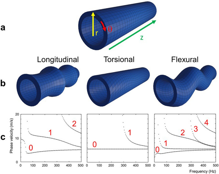
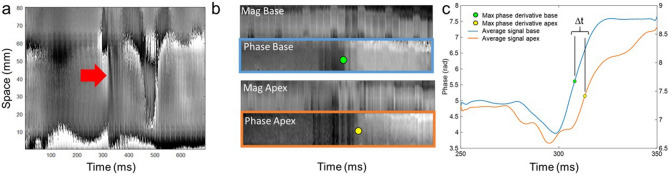
Changes in myocardial stiffness may represent a valuable biomarker for early tissue injury or adverse remodeling. In this study, we developed and validated a novel transducer-free magnetic resonance elastography (MRE) approach for quantifying myocardial biomechanics using aortic valve closure-induced shear waves. Using motion-sensitized two-dimensional pencil beams, septal shear waves were imaged at high temporal resolution. Shear wave speed was measured using time-of-flight of waves travelling between two pencil beams and corrected for geometrical biases. After validation in phantoms, results from twelve healthy volunteers and five cardiac patients (two left ventricular hypertrophy, two myocardial infarcts, and one without confirmed pathology) were obtained. Torsional shear wave speed in the phantom was 3.0 ± 0.1 m/s, corresponding with reference speeds of 2.8 ± 0.1 m/s. Geometrically-biased flexural shear wave speed was 1.9 ± 0.1 m/s, corresponding with simulation values of 2.0 m/s. Corrected septal shear wave speeds were significantly higher in patients than healthy volunteers [14.1 (11.0-15.8) m/s versus 3.6 (2.7-4.3) m/s, p = 0.001]. The interobserver 95%-limits-of-agreement in healthy volunteers were ± 1.3 m/s and interstudy 95%-limits-of-agreement - 0.7 to 1.2 m/s. In conclusion, myocardial shear wave speed can be measured using aortic valve closure-induced shear waves, with cardiac patients showing significantly higher shear wave speeds than healthy volunteers. This non-invasive measure may provide valuable insights into the pathophysiology of heart failure.
心肌僵硬度的变化可能代表早期组织损伤或不良重构的有价值的生物标志物。在这项研究中,我们开发并验证了一种使用主动脉瓣关闭引起的剪切波来量化心肌生物力学的新型无传感器磁共振弹性成像(MRE)方法。使用运动敏感的二维铅笔束,以高时间分辨率成像间隔层剪切波。使用在两条铅笔束之间传播的波的飞行时间测量剪切波速度,并校正几何偏差。在体模中验证后,从 12 名健康志愿者和 5 名心脏病患者(2 名左心室肥厚、2 名心肌梗死和 1 名无明确病理)获得了结果。在体模中扭转剪切波速度为 3.0±0.1 m/s,与参考速度 2.8±0.1 m/s 相对应。几何偏差弯曲剪切波速度为 1.9±0.1 m/s,与模拟值 2.0 m/s 相对应。患者的间隔层剪切波速度明显高于健康志愿者[14.1(11.0-15.8)m/s 与 3.6(2.7-4.3)m/s,p=0.001]。健康志愿者的观察者间 95%一致性界限为±1.3 m/s,研究间 95%一致性界限为-0.7 至 1.2 m/s。总之,使用主动脉瓣关闭引起的剪切波可以测量心肌剪切波速度,心脏病患者的剪切波速度明显高于健康志愿者。这种非侵入性测量方法可能为心力衰竭的病理生理学提供有价值的见解。