Hefei National Laboratory for Physical Sciences at Microscale, School of Life Sciences, National Synchrotron Radiation Laboratory, University of Science and Technology of China, Hefei, Anhui, China; Department of Cell Biology and Anatomy, Graduate School of Medicine, University of Tokyo, Tokyo, Japan.
Hefei National Laboratory for Physical Sciences at Microscale, School of Life Sciences, National Synchrotron Radiation Laboratory, University of Science and Technology of China, Hefei, Anhui, China.
J Biol Chem. 2020 Dec 18;295(51):17865-17876. doi: 10.1074/jbc.RA120.016295.
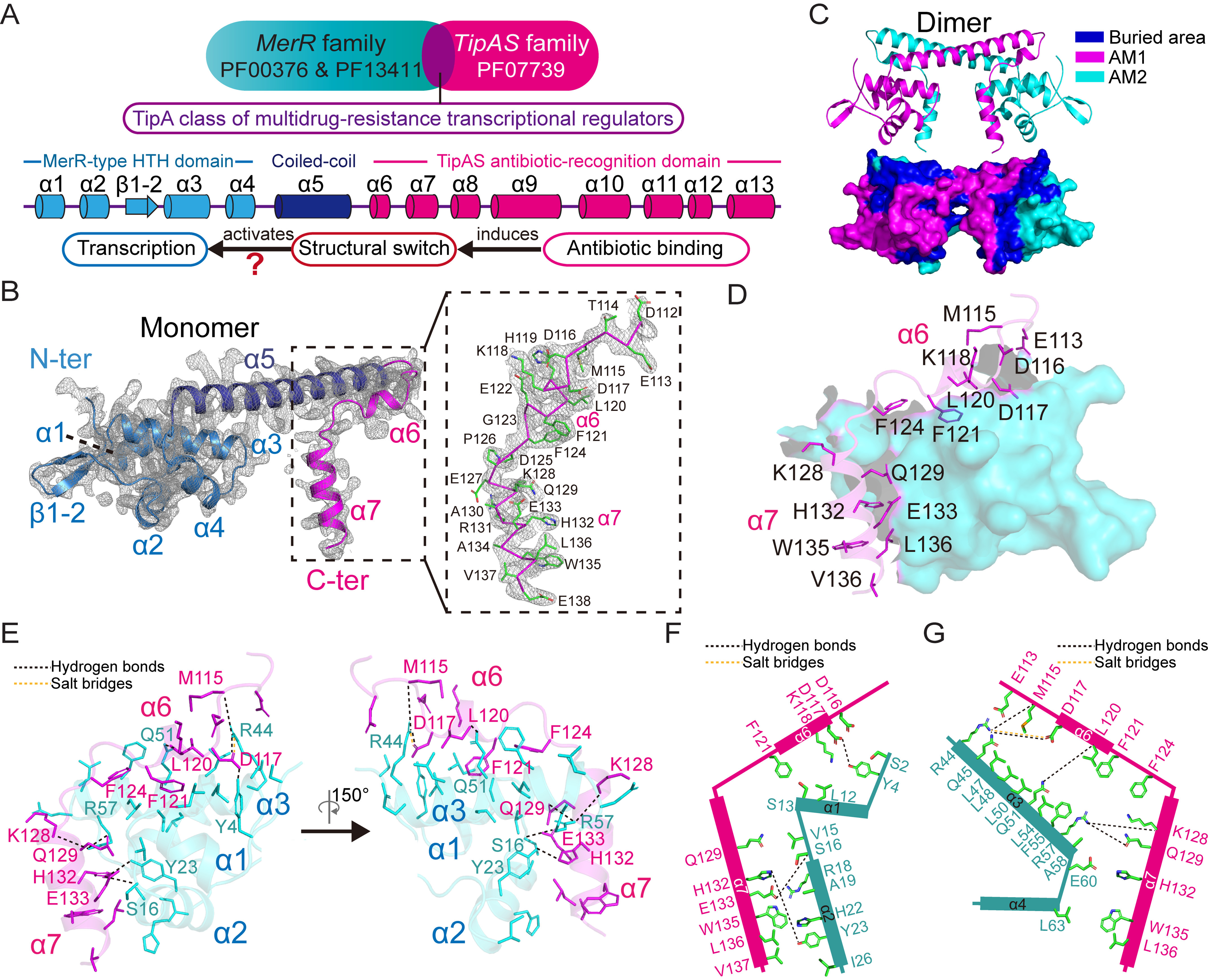
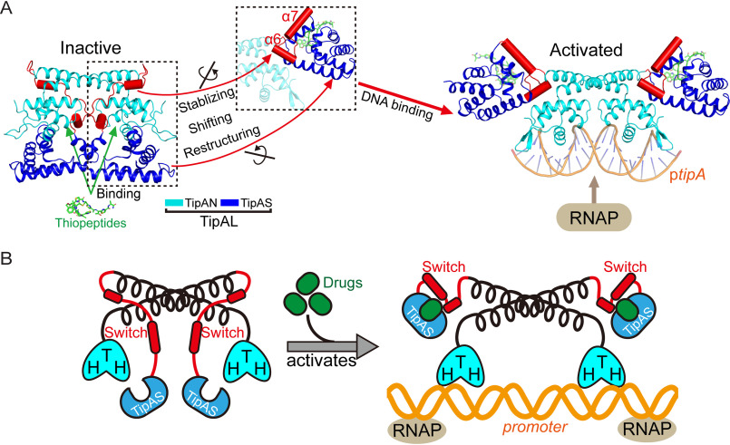
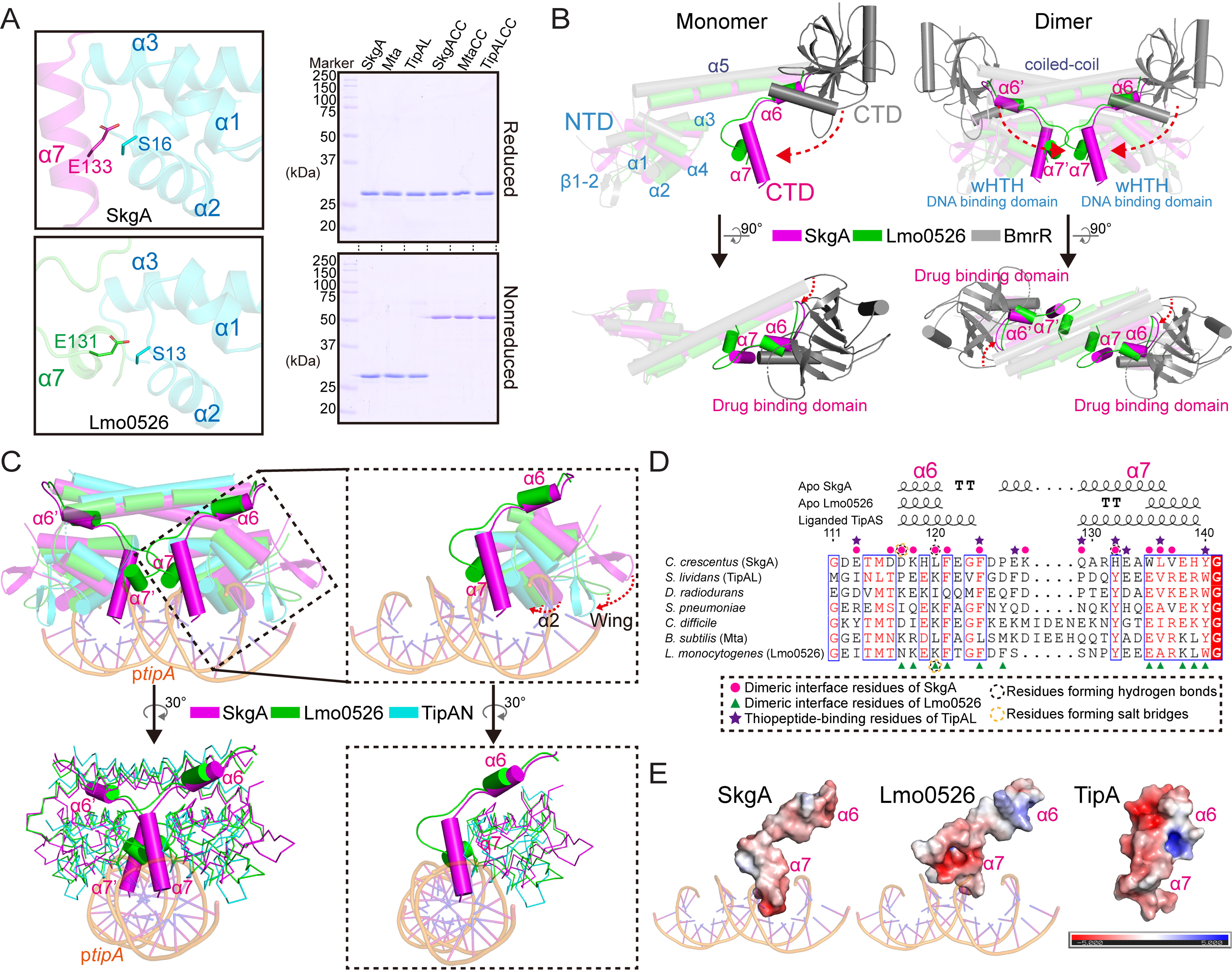
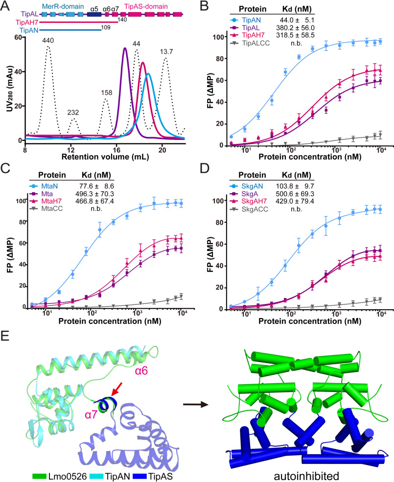
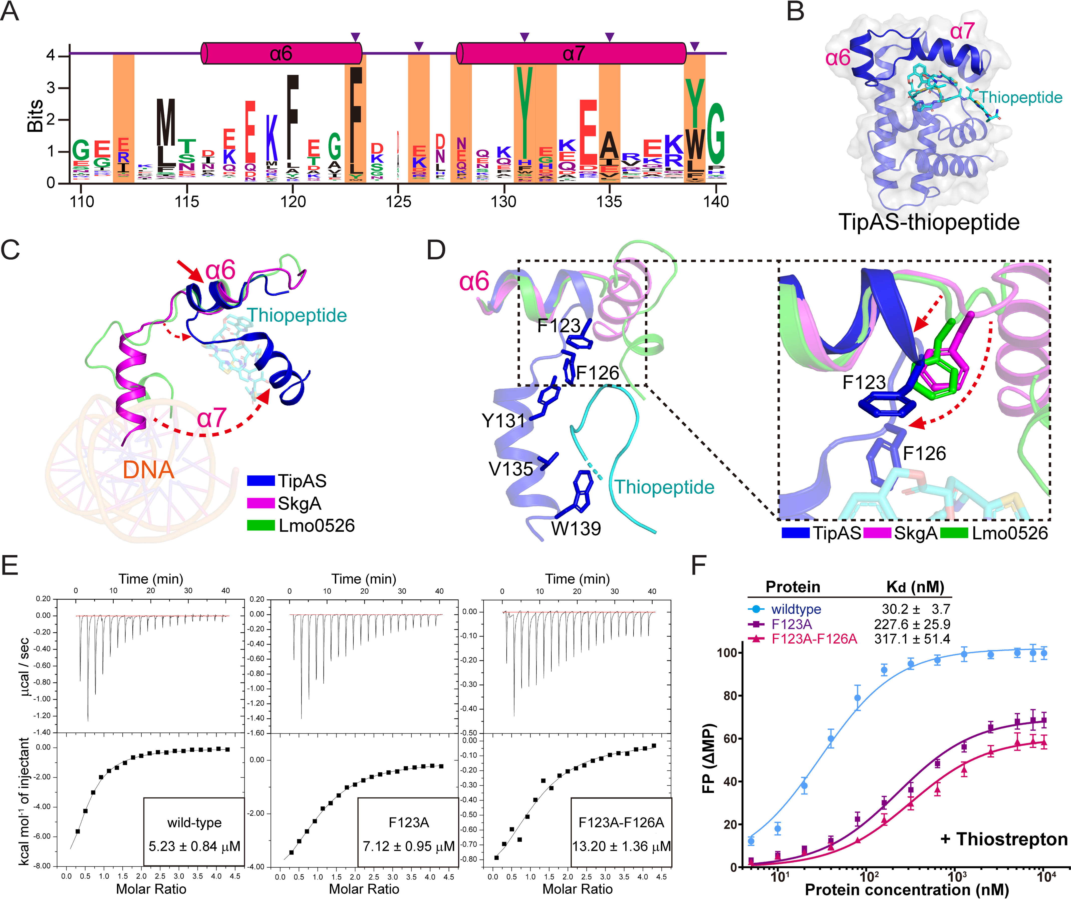
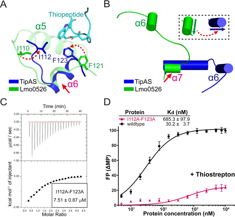
Investigations of bacterial resistance strategies can aid in the development of new antimicrobial drugs as a countermeasure to the increasing worldwide prevalence of bacterial antibiotic resistance. One such strategy involves the TipA class of transcription factors, which constitute minimal autoregulated multidrug resistance (MDR) systems against diverse antibiotics. However, we have insufficient information regarding how antibiotic binding induces transcriptional activation to design molecules that could interfere with this process. To learn more, we determined the crystal structure of SkgA from Caulobacter crescentus as a representative TipA protein. We identified an unexpected spatial orientation and location of the antibiotic-binding TipAS effector domain in the apo state. We observed that the α6-α7 region of the TipAS domain, which is canonically responsible for forming the lid of antibiotic-binding cleft to tightly enclose the bound antibiotic, is involved in the dimeric interface and stabilized via interaction with the DNA-binding domain in the apo state. Further structural and biochemical analyses demonstrated that the unliganded TipAS domain sterically hinders promoter DNA binding but undergoes a remarkable conformational shift upon antibiotic binding to release this autoinhibition via a switch of its α6-α7 region. Hence, the promoters for MDR genes including tipA and RNA polymerases become available for transcription, enabling efficient antibiotic resistance. These insights into the molecular mechanism of activation of TipA proteins advance our understanding of TipA proteins, as well as bacterial MDR systems, and may provide important clues to block bacterial resistance.
研究细菌耐药策略可以帮助开发新的抗菌药物,作为应对全球范围内细菌抗生素耐药性日益增加的对策。其中一种策略涉及 TipA 类转录因子,它们构成了针对多种抗生素的最小自我调节的多药耐药 (MDR) 系统。然而,我们对于抗生素结合如何诱导转录激活的信息还不够充分,无法设计出可以干扰这一过程的分子。为了了解更多信息,我们确定了新月柄杆菌 SkgA 作为 TipA 蛋白的代表的晶体结构。我们在无配体状态下发现了抗生素结合 TipAS 效应结构域的意外空间取向和位置。我们观察到,TipAS 结构域的 α6-α7 区域通常负责形成抗生素结合裂隙的盖子,以紧密包围结合的抗生素,该区域参与二聚体界面,并在无配体状态下通过与 DNA 结合结构域相互作用而稳定。进一步的结构和生化分析表明,未配体结合的 TipAS 结构域在空间上阻碍了启动子 DNA 结合,但在抗生素结合后会发生显著的构象变化,通过其 α6-α7 区域的转换释放这种自身抑制。因此,包括 tipA 和 RNA 聚合酶在内的 MDR 基因的启动子可用于转录,从而实现有效的抗生素耐药性。这些关于 TipA 蛋白激活的分子机制的见解,推进了我们对 TipA 蛋白以及细菌 MDR 系统的理解,并可能为阻止细菌耐药性提供重要线索。