• 文献检索
  • 文档翻译
  • 深度研究
  • 学术资讯
  • Suppr Zotero 插件Zotero 插件
  • 邀请有礼
  • 套餐&价格
  • 历史记录
应用&插件
Suppr Zotero 插件Zotero 插件浏览器插件Mac 客户端Windows 客户端微信小程序
定价
高级版会员购买积分包购买API积分包
服务
文献检索文档翻译深度研究API 文档MCP 服务
关于我们
关于 Suppr公司介绍联系我们用户协议隐私条款
关注我们

Suppr 超能文献

核心技术专利:CN118964589B侵权必究
粤ICP备2023148730 号-1Suppr @ 2026

文献检索

告别复杂PubMed语法,用中文像聊天一样搜索,搜遍4000万医学文献。AI智能推荐,让科研检索更轻松。

立即免费搜索

文件翻译

保留排版,准确专业,支持PDF/Word/PPT等文件格式,支持 12+语言互译。

免费翻译文档

深度研究

AI帮你快速写综述,25分钟生成高质量综述,智能提取关键信息,辅助科研写作。

立即免费体验

通过与自动分割的F-氟脱氧葡萄糖正电子发射断层扫描相比较,评估扩散加权成像在头颈部放射治疗中肿瘤轮廓描绘的作用。

Evaluation of diffusion weighted imaging for tumor delineation in head-and-neck radiotherapy by comparison with automatically segmented F-fluorodeoxyglucose positron emission tomography.

作者信息

Schakel Tim, Peltenburg Boris, Dankbaar Jan-Willem, Cardenas Carlos E, Aristophanous Michalis, Terhaard Chris H J, Hoogduin Johannes M, Philippens Marielle E P

机构信息

Department of Radiotherapy, University Medical Center, Utrecht, The Netherlands.

Department of Radiology, University Medical Center, Utrecht, The Netherlands.

出版信息

Phys Imaging Radiat Oncol. 2018 Jan 30;5:13-18. doi: 10.1016/j.phro.2017.12.004. eCollection 2018 Jan.

DOI:10.1016/j.phro.2017.12.004
PMID:33458363
原文链接:https://pmc.ncbi.nlm.nih.gov/articles/PMC7807628/
Abstract

BACKGROUND AND PURPOSE

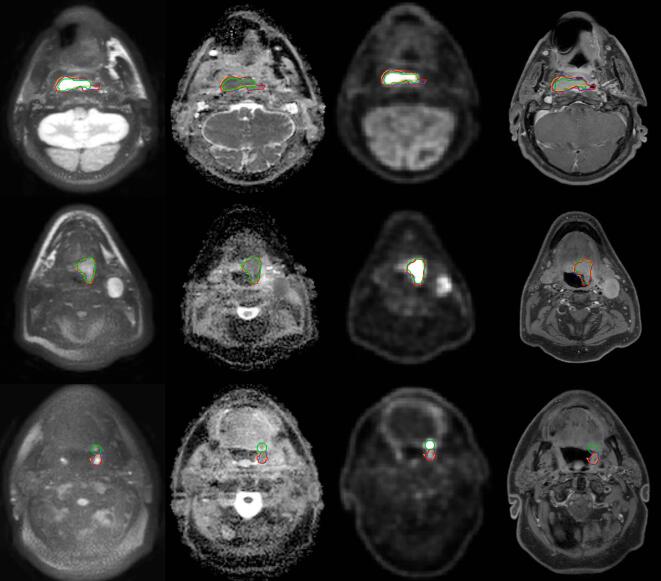
Diffusion weighted (DW) MRI may facilitate target volume delineation for head-and-neck (HN) radiation treatment planning. In this study we assessed the use of a dedicated, geometrically accurate, DW-MRI sequence for target volume delineation. The delineations were compared with semi-automatic segmentations on F-fluorodeoxyglucose (FDG) positron emission tomography (PET) images and evaluated for interobserver variation.

METHODS AND MATERIALS

Fifteen HN cancer patients underwent both DW-MRI and FDG-PET for RT treatment planning. Target delineation on DW-MRI was performed by three observers, while for PET a semi-automatic segmentation was performed using a Gaussian mixture model. For interobserver variation and intermodality variation, volumes, overlap metrics and Hausdorff distances were calculated from the delineations.

RESULTS

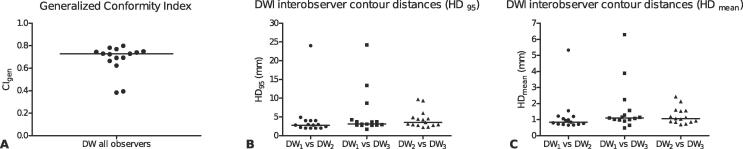
The median volumes delineated by the three observers on DW-MRI were 10.8, 10.5 and 9.0 cm respectively, and was larger than the median PET volume (8.0 cm). The median conformity index of DW-MRI for interobserver variation was 0.73 (range 0.38-0.80). Compared to PET, the delineations on DW-MRI by the three observers showed a median dice similarity coefficient of 0.71, 0.69 and 0.72 respectively. The mean Hausdorff distance was small with median (range) distances between PET and DW-MRI of 2.3 (1.5-6.8), 2.5 (1.6-6.9) and 2.0 (1.35-7.6) mm respectively. Over all patients, the median 95th percentile distances were 6.0 (3.0-13.4), 6.6 (4.0-24.0) and 5.3 (3.4-26.0) mm.

CONCLUSION

Using a dedicated DW-MRI sequence, target volumes could be defined with good interobserver agreement and a good overlap with PET. Target volume delineation using DW-MRI is promising in head-and-neck radiotherapy, combined with other modalities, it can lead to more precise target volume delineation.

摘要

背景与目的

扩散加权磁共振成像(DW-MRI)有助于对头颈部(HN)放射治疗计划中的靶区进行勾画。在本研究中,我们评估了一种专门的、几何精度高的DW-MRI序列用于靶区勾画的情况。将这些勾画结果与基于氟脱氧葡萄糖(FDG)正电子发射断层扫描(PET)图像的半自动分割结果进行比较,并评估观察者间的差异。

方法与材料

15例HN癌症患者同时接受了DW-MRI和FDG-PET检查以进行放疗计划。三位观察者对DW-MRI图像进行靶区勾画,而对于PET图像,则使用高斯混合模型进行半自动分割。从勾画结果中计算观察者间差异和模态间差异的体积、重叠指标和豪斯多夫距离。

结果

三位观察者在DW-MRI上勾画的中位体积分别为10.8、10.5和9.0 cm³,大于PET的中位体积(8.0 cm³)。观察者间差异的DW-MRI中位符合指数为0.73(范围0.38 - 0.80)。与PET相比,三位观察者在DW-MRI上的勾画结果的中位骰子相似系数分别为0.71、0.69和0.72。平均豪斯多夫距离较小,PET与DW-MRI之间距离的中位数(范围)分别为2.3(1.5 - 6.8)、2.5(1.6 - 6.9)和2.0(1.35 - 7.6)mm。在所有患者中,第95百分位数距离的中位数分别为6.0(3.0 - 13.4)、6.6(4.0 - 24.0)和5.3(3.4 - 26.0)mm。

结论

使用专门的DW-MRI序列,可以在观察者间达成良好的一致性且与PET有良好的重叠来定义靶区体积。在头颈部放疗中,使用DW-MRI进行靶区勾画很有前景,与其他模态相结合,可实现更精确的靶区勾画。

https://cdn.ncbi.nlm.nih.gov/pmc/blobs/460d/7807628/66090888bf2a/gr4.jpg
https://cdn.ncbi.nlm.nih.gov/pmc/blobs/460d/7807628/a274be555201/gr1.jpg
https://cdn.ncbi.nlm.nih.gov/pmc/blobs/460d/7807628/84518e954799/gr2.jpg
https://cdn.ncbi.nlm.nih.gov/pmc/blobs/460d/7807628/42238d585392/gr3.jpg
https://cdn.ncbi.nlm.nih.gov/pmc/blobs/460d/7807628/66090888bf2a/gr4.jpg
https://cdn.ncbi.nlm.nih.gov/pmc/blobs/460d/7807628/a274be555201/gr1.jpg
https://cdn.ncbi.nlm.nih.gov/pmc/blobs/460d/7807628/84518e954799/gr2.jpg
https://cdn.ncbi.nlm.nih.gov/pmc/blobs/460d/7807628/42238d585392/gr3.jpg
https://cdn.ncbi.nlm.nih.gov/pmc/blobs/460d/7807628/66090888bf2a/gr4.jpg

相似文献

1
Evaluation of diffusion weighted imaging for tumor delineation in head-and-neck radiotherapy by comparison with automatically segmented F-fluorodeoxyglucose positron emission tomography.通过与自动分割的F-氟脱氧葡萄糖正电子发射断层扫描相比较,评估扩散加权成像在头颈部放射治疗中肿瘤轮廓描绘的作用。
Phys Imaging Radiat Oncol. 2018 Jan 30;5:13-18. doi: 10.1016/j.phro.2017.12.004. eCollection 2018 Jan.
2
Gross Tumor Delineation in Esophageal Cancer on MRI Compared With F-FDG-PET/CT.食管癌MRI与F-FDG-PET/CT的大体肿瘤勾画比较
Adv Radiat Oncol. 2019 Apr 24;4(4):596-604. doi: 10.1016/j.adro.2019.04.004. eCollection 2019 Oct-Dec.
3
Geometrical Comparison and Quantitative Evaluation of F-FDG PET/CT- and DW-MRI-Based Target Delineation Before and During Radiotherapy for Esophageal Squamous Carcinoma.基于F-FDG PET/CT和扩散加权磁共振成像的食管癌放疗前及放疗期间靶区勾画的几何比较与定量评估
Front Oncol. 2021 Dec 22;11:772428. doi: 10.3389/fonc.2021.772428. eCollection 2021.
4
Evaluating diffusion-weighted magnetic resonance imaging for target volume delineation in head and neck radiotherapy.评估扩散加权磁共振成像在头颈部放射治疗中靶区勾画的应用
J Med Imaging Radiat Oncol. 2019 Jun;63(3):399-407. doi: 10.1111/1754-9485.12866. Epub 2019 Feb 28.
5
Deep learning-based automatic delineation of anal cancer gross tumour volume: a multimodality comparison of CT, PET and MRI.基于深度学习的肛门癌大体肿瘤体积自动勾画:CT、PET 和 MRI 的多模态比较。
Acta Oncol. 2022 Jan;61(1):89-96. doi: 10.1080/0284186X.2021.1994645. Epub 2021 Nov 16.
6
Variability of gross tumour volume delineation: MRI and CT based tumour and lymph node delineation for lung radiotherapy.大体肿瘤体积勾画的可变性:用于肺放疗的 MRI 和 CT 肿瘤及淋巴结勾画。
Radiother Oncol. 2022 Feb;167:292-299. doi: 10.1016/j.radonc.2021.11.036. Epub 2021 Dec 8.
7
Prostate cancer GTV delineation with biparametric MRI and Ga-PSMA-PET: comparison of expert contours and semi-automated methods.基于双参数 MRI 和 Ga-PSMA-PET 的前列腺癌 GTV 勾画:专家勾画与半自动勾画方法的比较。
Br J Radiol. 2021 Mar 1;94(1119):20201174. doi: 10.1259/bjr.20201174. Epub 2021 Jan 28.
8
A multimodality segmentation framework for automatic target delineation in head and neck radiotherapy.一种用于头颈部放射治疗中自动靶区勾画的多模态分割框架。
Med Phys. 2015 Sep;42(9):5310-20. doi: 10.1118/1.4928485.
9
Manual and semi-automated delineation of locally advanced rectal cancer subvolumes with diffusion-weighted MRI.基于弥散加权 MRI 的局部进展期直肠癌亚体积手动和半自动勾画。
Br J Radiol. 2020 Oct 1;93(1114):20200543. doi: 10.1259/bjr.20200543. Epub 2020 Sep 2.
10
Interobserver and intermodality variability in GTV delineation on simulation CT, FDG-PET, and MR Images of Head and Neck Cancer.头颈部癌模拟CT、FDG-PET和MR图像上大体肿瘤体积(GTV)勾画的观察者间和模态间变异性
Jacobs J Radiat Oncol. 2014 Sep;1(1):006.

引用本文的文献

1
The Use of MR-Guided Radiation Therapy for Head and Neck Cancer and Recommended Reporting Guidance.MR 引导放疗在头颈部肿瘤中的应用及推荐报告指南。
Semin Radiat Oncol. 2024 Jan;34(1):69-83. doi: 10.1016/j.semradonc.2023.10.003.
2
Prospective evaluation of in vivo and phantom repeatability and reproducibility of diffusion-weighted MRI sequences on 1.5 T MRI-linear accelerator (MR-Linac) and MR simulator devices for head and neck cancers.1.5T MRI 线性加速器(MR-Linac)和 MR 模拟器设备上扩散加权 MRI 序列在头颈部癌症中的体内和体模重复性及可重复性的前瞻性评估。
Radiother Oncol. 2023 Aug;185:109717. doi: 10.1016/j.radonc.2023.109717. Epub 2023 May 19.
3

本文引用的文献

1
Technical Note: Diffusion-weighted MRI with minimal distortion in head-and-neck radiotherapy using a turbo spin echo acquisition method.技术注释:使用涡轮自旋回波采集方法在头颈部放疗中实现最小失真的弥散加权 MRI。
Med Phys. 2017 Aug;44(8):4188-4193. doi: 10.1002/mp.12363. Epub 2017 Jun 30.
2
Inadequate target volume delineation and local-regional recurrence after intensity-modulated radiotherapy for human papillomavirus-positive oropharynx cancer.调强放疗后 HPV 阳性口咽癌靶区勾画不足与局部区域复发。
Radiother Oncol. 2017 Jun;123(3):412-418. doi: 10.1016/j.radonc.2017.04.015. Epub 2017 May 13.
3
Modality-specific target definition for laryngeal and hypopharyngeal cancer on FDG-PET, CT and MRI.
Optimized flip angle schemes for the split acquisition of fast spin-echo signals (SPLICE) sequence and application to diffusion-weighted imaging.
优化的翻转角方案用于快速自旋回波信号的分离采集(SPLICE 序列)及其在扩散加权成像中的应用。
Magn Reson Med. 2023 Apr;89(4):1469-1480. doi: 10.1002/mrm.29545. Epub 2022 Nov 24.
4
Target Definition in MR-Guided Adaptive Radiotherapy for Head and Neck Cancer.头颈部癌磁共振引导自适应放射治疗中的靶区定义
Cancers (Basel). 2022 Jun 20;14(12):3027. doi: 10.3390/cancers14123027.
5
Effects of different CT value assignment methods on dose calculations in radiotherapy planning for brain metastases.不同CT值赋值方法对脑转移瘤放射治疗计划剂量计算的影响。
Transl Cancer Res. 2020 Feb;9(2):1091-1099. doi: 10.21037/tcr.2019.12.54.
6
Early Response to Chemotherapy in Malignant Pleural Mesothelioma Evaluated Using Diffusion-Weighted Magnetic Resonance Imaging: Initial Observations.使用扩散加权磁共振成像评估恶性胸膜间皮瘤对化疗的早期反应:初步观察
JTO Clin Res Rep. 2021 Nov 2;2(12):100253. doi: 10.1016/j.jtocrr.2021.100253. eCollection 2021 Dec.
7
Imaging science and development in modern high-precision radiotherapy.现代高精度放射治疗中的影像科学与发展
Phys Imaging Radiat Oncol. 2019 Dec 9;12:63-66. doi: 10.1016/j.phro.2019.11.008. eCollection 2019 Oct.
8
Recent advances in MRI of the head and neck, skull base and cranial nerves: new and evolving sequences, analyses and clinical applications.头颈部、颅底和颅神经 MRI 的最新进展:新的和不断发展的序列、分析和临床应用。
Br J Radiol. 2019 Dec;92(1104):20190513. doi: 10.1259/bjr.20190513. Epub 2019 Sep 24.
基于氟代脱氧葡萄糖正电子发射断层扫描(FDG-PET)、计算机断层扫描(CT)和磁共振成像(MRI)的喉癌和下咽癌的模态特异性靶区定义
Radiother Oncol. 2017 Apr;123(1):63-70. doi: 10.1016/j.radonc.2017.02.005. Epub 2017 Mar 1.
4
Simultaneous (18)F-FDG-PET/MRI: Associations between diffusion, glucose metabolism and histopathological parameters in patients with head and neck squamous cell carcinoma.同步(18)F-FDG-PET/MRI:头颈部鳞状细胞癌患者扩散、葡萄糖代谢与组织病理学参数之间的关联
Oral Oncol. 2016 Jul;58:14-20. doi: 10.1016/j.oraloncology.2016.04.009. Epub 2016 May 7.
5
Radiation oncology in the era of precision medicine.精准医学时代的放射肿瘤学。
Nat Rev Cancer. 2016 Apr;16(4):234-49. doi: 10.1038/nrc.2016.18. Epub 2016 Mar 18.
6
Inter-observer agreement of MRI-based tumor delineation for preoperative radiotherapy boost in locally advanced rectal cancer.基于 MRI 的局部晚期直肠癌术前放疗增敏的肿瘤勾画的观察者间一致性。
Radiother Oncol. 2016 Feb;118(2):399-407. doi: 10.1016/j.radonc.2015.10.030. Epub 2015 Dec 14.
7
Diffusion-weighted magnetic resonance imaging for the prediction of pathologic response to neoadjuvant chemoradiotherapy in esophageal cancer.扩散加权磁共振成像预测食管癌新辅助放化疗的病理反应
Radiother Oncol. 2015 May;115(2):163-70. doi: 10.1016/j.radonc.2015.04.027. Epub 2015 May 19.
8
Alterations in anatomic and functional imaging parameters with repeated FDG PET-CT and MRI during radiotherapy for head and neck cancer: a pilot study.头颈部癌放疗期间重复进行氟代脱氧葡萄糖正电子发射断层扫描-计算机断层扫描(FDG PET-CT)和磁共振成像(MRI)时解剖和功能成像参数的变化:一项初步研究
BMC Cancer. 2015 Mar 17;15:137. doi: 10.1186/s12885-015-1154-8.
9
The accuracy of target delineation in laryngeal and hypopharyngeal cancer.喉癌和下咽癌中靶区勾画的准确性。
Acta Oncol. 2015;54(8):1181-7. doi: 10.3109/0284186X.2015.1006401. Epub 2015 Mar 3.
10
Response evaluation after chemoradiotherapy for advanced nodal disease in head and neck cancer using diffusion-weighted MRI and 18F-FDG-PET-CT.使用扩散加权磁共振成像(MRI)和18F-氟代脱氧葡萄糖正电子发射断层扫描-计算机断层扫描(18F-FDG-PET-CT)对头颈部癌晚期淋巴结疾病进行放化疗后的疗效评估
Oral Oncol. 2015 May;51(5):541-7. doi: 10.1016/j.oraloncology.2015.01.017. Epub 2015 Feb 26.