Taasti Vicki Trier, Muren Ludvig Paul, Jensen Kenneth, Petersen Jørgen Breede Baltzer, Thygesen Jesper, Tietze Anna, Grau Cai, Hansen David Christoffer
Dept. of Medical Physics, Aarhus University Hospital, Aarhus, Denmark.
Dept. of Oncology, Aarhus University Hospital, Aarhus, Denmark.
Phys Imaging Radiat Oncol. 2018 Apr 22;6:14-19. doi: 10.1016/j.phro.2018.04.002. eCollection 2018 Apr.
Patients with head and neck (HN) cancer may benefit from proton therapy due to the potential for sparing of normal tissue. For planning of proton therapy, dual-energy CT (DECT) has been shown to provide superior stopping power ratio (SPR) determination in phantom materials and organic tissue samples, compared to single-energy CT (SECT). However, the benefit of DECT in HN cancer patients has not yet been investigated. This study therefore compared DECT- and SECT-based SPR estimation for HN cancer patients.
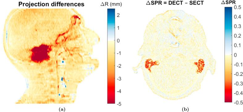
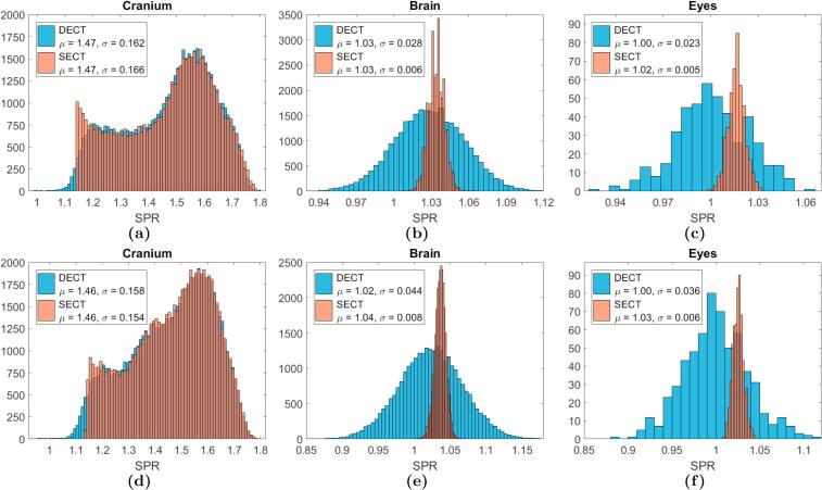
Fourteen HN cancer patients were DECT scanned. Eight patients were scanned using a dual source DECT scanner and six were scanned with a conventional SECT scanner by acquiring two consecutive scans. SECT image sets were computed as a weighted summation of the low and high energy DECT image sets. DECT- and SECT-based SPR maps were derived. Water-equivalent path lengths (WEPLs) through the SPR maps were compared in the eight cases with dual source DECT scans. Mean SPR estimates over region-of-interests (ROIs) in the cranium, brain and eyes were analyzed for all patients.
A median WEPL difference of 1.9 mm (1.5%) was found across the eight patients. Statistically significant SPR differences were seen for the ROIs in the brain and eyes, with the SPR estimates based on DECT overall lower than for SECT.
Clinically relevant WEPL and SPR differences were found between DECT and SECT, which could imply that the accuracy of treatment planning for proton therapy would benefit from DECT-based SPR estimation.
头颈部(HN)癌患者可能因质子治疗对正常组织的潜在保护作用而受益。在质子治疗计划中,与单能CT(SECT)相比,双能CT(DECT)已被证明在体模材料和有机组织样本中能提供更优的阻止本领比(SPR)测定。然而,DECT在HN癌患者中的益处尚未得到研究。因此,本研究比较了基于DECT和SECT的HN癌患者SPR估计值。
对14例HN癌患者进行了DECT扫描。8例患者使用双源DECT扫描仪进行扫描,6例患者通过连续采集两次扫描,使用传统的SECT扫描仪进行扫描。SECT图像集通过低能和高能DECT图像集的加权求和计算得出。得出基于DECT和SECT的SPR图。在8例使用双源DECT扫描的病例中,比较了通过SPR图的水等效路径长度(WEPL)。分析了所有患者颅骨、脑和眼中感兴趣区域(ROI)的平均SPR估计值。
8例患者的WEPL中位数差异为1.9毫米(1.5%)。在脑和眼的ROI中观察到SPR有统计学显著差异,基于DECT的SPR估计总体低于SECT。
在DECT和SECT之间发现了具有临床相关性的WEPL和SPR差异,这可能意味着基于DECT的SPR估计将有益于质子治疗计划的准确性。