Valstar Matthijs H, Owers Emilia C, Al-Mamgani Abrahim, Smeele Ludwig E, van de Kamer Jeroen B, Sonke Jan-Jakob, Vogel Wouter V
Department of Head and Neck Oncology and Surgery, Antoni van Leeuwenhoek The Netherlands Cancer Institute, Amsterdam, the Netherlands.
Department of Oral and Maxillofacial Surgery, Amsterdam UMC, University of Amsterdam, the Netherlands.
Phys Imaging Radiat Oncol. 2019 Mar 4;9:65-68. doi: 10.1016/j.phro.2019.02.004. eCollection 2019 Jan.
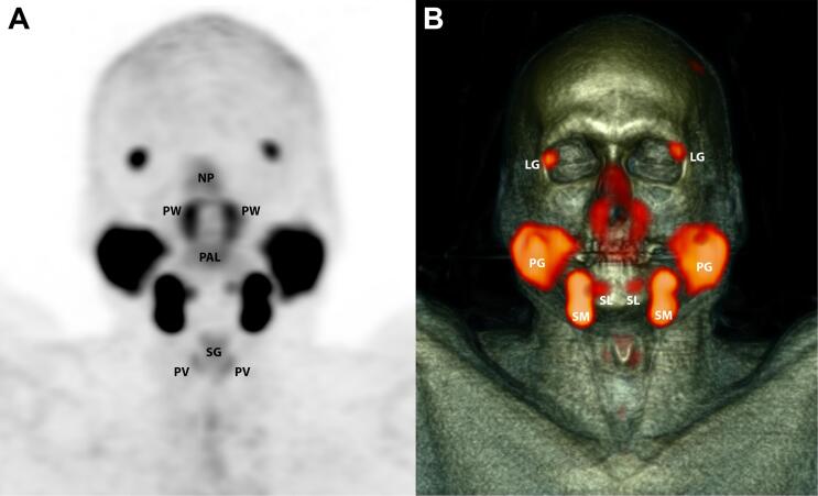
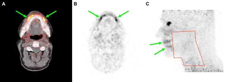
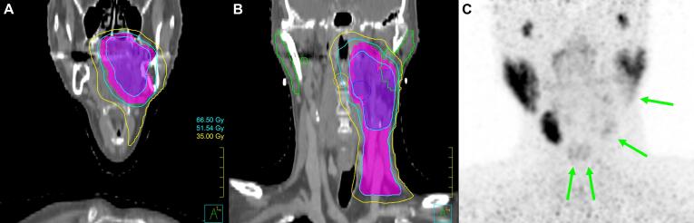
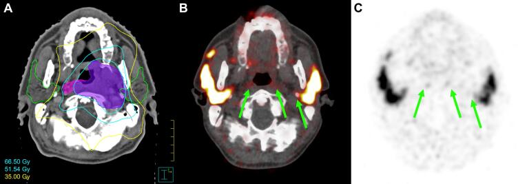
Evaluation of salivary gland damage after head and neck radiotherapy (RT) is difficult with current tools, such as subjective patient-reported outcome measures. We demonstrate the use of prostate-specific membrane antigen positron emission tomography/computed tomography (PSMA PET/CT) as an objective non-invasive tool to visualize damage to salivary glands resulting from RT. In three clinical cases, the PSMA-ligand distribution correlates to the RT dose distribution including intra-gland dose gradients and matches patient-reported toxicity, suggesting a dose-response relation. These findings support further exploration of PSMA PET/CT to guide and evaluate RT, with the ultimate aim to reduce salivary gland toxicity.
使用当前工具(如患者主观报告的结局指标)评估头颈部放疗(RT)后的唾液腺损伤存在困难。我们展示了使用前列腺特异性膜抗原正电子发射断层扫描/计算机断层扫描(PSMA PET/CT)作为一种客观的非侵入性工具,以可视化放疗导致的唾液腺损伤。在三个临床病例中,PSMA配体分布与放疗剂量分布相关,包括腺体内剂量梯度,并与患者报告的毒性相匹配,提示存在剂量反应关系。这些发现支持进一步探索PSMA PET/CT以指导和评估放疗,最终目标是降低唾液腺毒性。