Faculty of Health and Medicine, University of Newcastle, Newcastle, Australia.
NHMRC Center of Research Excellence in Digestive Health Newcastle, Australia.
Clin Transl Gastroenterol. 2020 Dec 22;12(1):e00296. doi: 10.14309/ctg.0000000000000296.
Histopathological alterations in the ileum and colon in irritable bowel syndrome (IBS) are controversial, and normal values are poorly established. We hypothesized that changes in mucosal immune cells characterize IBS and key changes in immune composition are associated with the mucosa-associated microbiota (MaM).
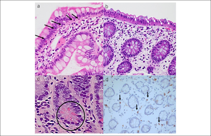
A nested case-control study (48 IBS and 106 controls included) from 745 colonoscopy participants in a random population sample. Intraepithelial lymphocytes (IELs)/100 enterocytes and eosinophils/5 nonoverlapping high-power fields counted; mast cells identified by immunocytochemistry (CD117)/5 high-power fields. Paneth cells quantified per 5 crypts. 16S rRNA gene amplicon sequencing performed on available sigmoid MaM, n = 55 and fecal microbiota, n = 20. Microbiota profiles compared between samples with high and low IEL counts.
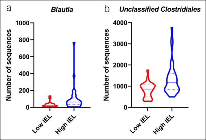
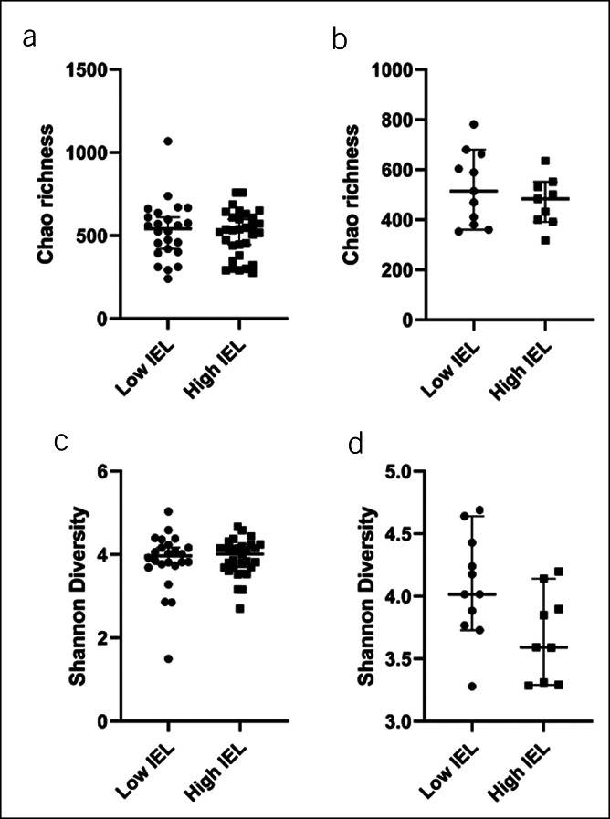
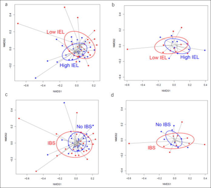
IBS had increased IELs in the terminal ileum (relative risk ratio = 1.70, 95% confidence interval 1.08-2.76, P = 0.022 adjusted for age, sex, and smoking). Cecal IELs were increased in IBS-diarrhea (relative risk ratio = 2.03, 95% confidence interval 1.13-3.63, P = 0.017). No difference was observed in alpha diversity of MaM or fecal microbiota based on IEL count. There was no difference in beta diversity of the MaM according to IEL count in the terminal ileal (TI) (P = 0.079). High TI IEL counts associated with a significant expansion of the genus Blautia (P = 0.024) and unclassified Clostridiales (P = 0.036) in colon MaM.
A modest but significant increase in IELs was observed in IBS vs. controls in a population-based setting. Subtle TI and cecal inflammation may play a pathogenic role in IBS but needs confirmation. Modest but discernible differences in the colonic MaM were seen according to TI IEL count but not IBS status.
在肠易激综合征(IBS)中,回肠和结肠的组织病理学改变存在争议,且正常参考值尚未明确。我们假设黏膜免疫细胞的变化可作为 IBS 的特征,而免疫组成的关键变化与黏膜相关微生物群(MaM)有关。
在一项随机人群样本的结肠镜检查参与者中(共纳入 745 例,其中 48 例为 IBS 患者,106 例为对照组),我们进行了嵌套病例对照研究。计数 100 个肠上皮细胞中每 100 个的固有层淋巴细胞(IELs)和嗜酸性粒细胞/5 个不重叠的高倍视野;通过免疫细胞化学鉴定 5 个高倍视野中的肥大细胞(CD117)。每个 5 个隐窝计算潘氏细胞的数量。对 55 例可获得的乙状结肠 MaM 和 20 例粪便微生物群进行 16S rRNA 基因扩增子测序。比较 IEL 计数高和低的样本之间的微生物组谱。
IBS 患者的末端回肠 IELs 增加(相对风险比=1.70,95%置信区间 1.08-2.76,P=0.022,调整了年龄、性别和吸烟因素)。IBS-腹泻患者的盲肠 IELs 增加(相对风险比=2.03,95%置信区间 1.13-3.63,P=0.017)。根据 IEL 计数,MaM 或粪便微生物群的 alpha 多样性没有差异。根据 IEL 计数,末端回肠(TI)的 MaM 的 beta 多样性没有差异(P=0.079)。高 TI IEL 计数与结肠 MaM 中明显扩张的布劳特氏菌属(P=0.024)和未分类的梭状芽胞杆菌属(P=0.036)相关。
在人群中,我们观察到 IBS 患者与对照组相比,IELs 有适度但显著的增加。细微的 TI 和盲肠炎症可能在 IBS 中发挥致病作用,但需要进一步证实。根据 TI IEL 计数,结肠 MaM 存在适度但可识别的差异,但与 IBS 状态无关。