Department of Medical Imaging, Radboud University Medical Center, Geert Grooteplein 10, 6525, GA, Nijmegen, The Netherlands.
Dutch Expert Centre for Screening (LRCB), Wijchenseweg 101, 6538, SW, Nijmegen, The Netherlands.
Eur Radiol. 2021 Jul;31(7):5335-5343. doi: 10.1007/s00330-020-07679-8. Epub 2021 Jan 21.
To study how radiologists' perceived ability to interpret digital mammography (DM) images is affected by decreases in image quality.
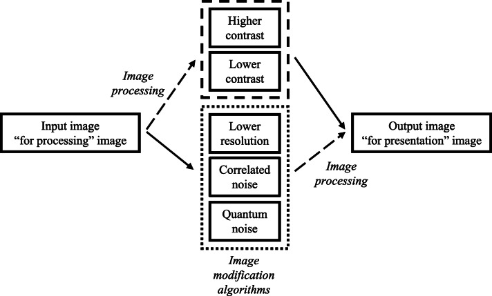
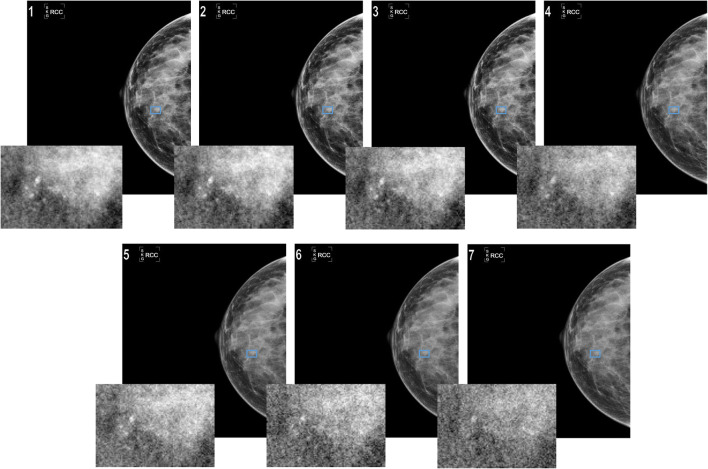
One view from 45 DM cases (including 30 cancers) was degraded to six levels each of two acquisition-related issues (lower spatial resolution and increased quantum noise) and three post-processing-related issues (lower and higher contrast and increased correlated noise) seen during clinical evaluation of DM systems. The images were shown to fifteen breast screening radiologists from five countries. Aware of lesion location, the radiologists selected the most-degraded mammogram (indexed from 1 (reference) to 7 (most degraded)) they still felt was acceptable for interpretation. The median selected index, per degradation type, was calculated separately for calcification and soft tissue (including normal) cases. Using the two-sided, non-parametric Mann-Whitney test, the median indices for each case and degradation type were compared.
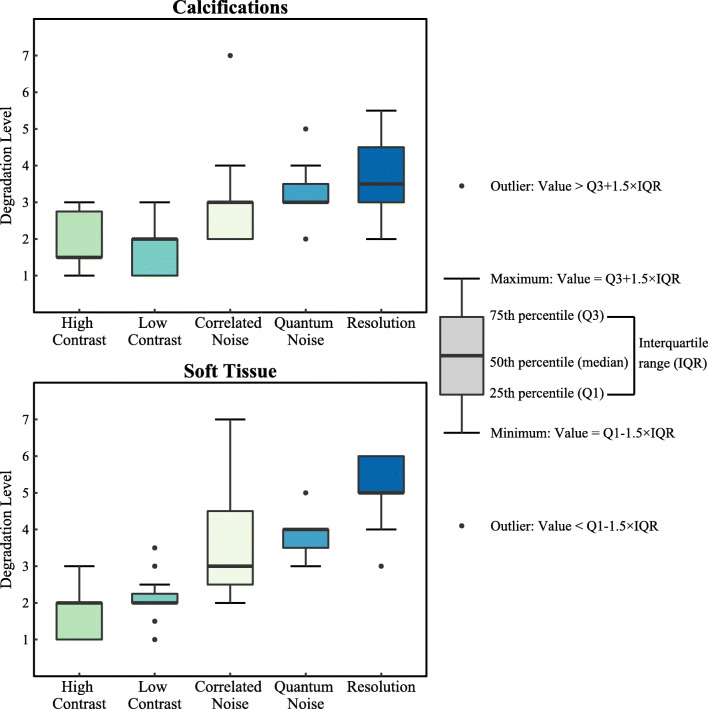
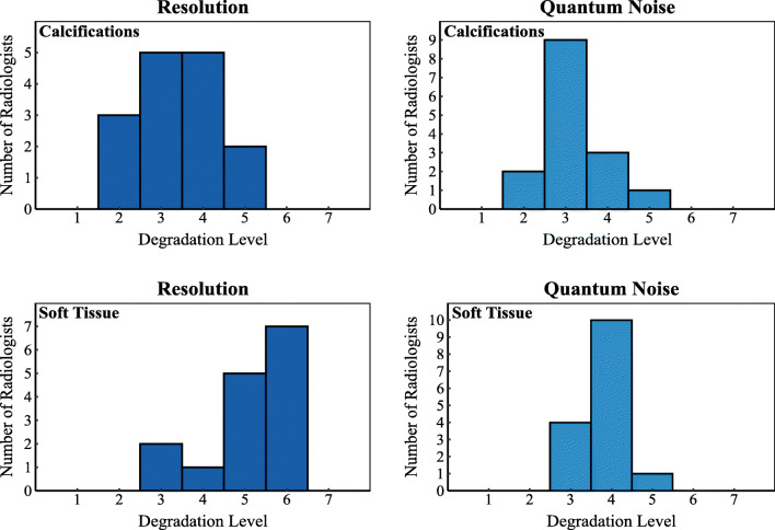
Radiologists were not tolerant to increases (medians: 1.5 (calcifications) and 2 (soft tissue)) or decreases (median: 2, for both types) in contrast, but were more tolerant to correlated noise (median: 3, for both types). Increases in quantum noise were tolerated more for calcifications than for soft tissue cases (medians: 3 vs. 4, p = 0.02). Spatial resolution losses were considered less acceptable for calcification detection than for soft tissue cases (medians: 3.5 vs. 5, p = 0.001).
Perceived ability of radiologists for image interpretation in DM was affected not only by image acquisition-related issues but also by image post-processing issues, and some of those issues affected calcification cases more than soft tissue cases.
• Lower spatial resolution and increased quantum noise affected the radiologists' perceived ability to interpret calcification cases more than soft tissue lesion or normal cases. • Post-acquisition image processing-related effects, not only image acquisition-related effects, also impact the perceived ability of radiologists to interpret images and detect lesions. • In addition to current practices, post-acquisition image processing-related effects need to also be considered during the testing and evaluation of digital mammography systems.
研究放射科医生对数字乳腺 X 线摄影(DM)图像的解读能力如何受到图像质量下降的影响。
从 45 例 DM 病例(包括 30 例癌症)中选择一个视图,该视图的两种与获取相关的问题(空间分辨率降低和量子噪声增加)和三种与后处理相关的问题(对比度降低和增加、相关噪声增加)各降低六个等级,这些问题是在 DM 系统的临床评估中观察到的。将这些图像展示给来自五个国家的 15 名乳腺筛查放射科医生。这些放射科医生在了解病变位置的情况下,选择他们认为仍然可以接受解释的最严重退化的乳腺 X 线照片(从 1(参考)到 7(最严重)进行索引)。分别计算每种退化类型的钙化和软组织(包括正常)病例的中位数所选索引。使用双侧非参数曼-惠特尼检验,比较每种病例和退化类型的中位数指数。
放射科医生对对比度的增加(中位数:1.5(钙化)和 2(软组织))或降低(中位数:2,两种类型)都不宽容,但对相关噪声的容忍度更高(中位数:3,两种类型)。与软组织病例相比,对量子噪声的增加,钙化病例的容忍度更高(中位数:3 比 4,p = 0.02)。空间分辨率的损失被认为对钙化检测的可接受性低于软组织病例(中位数:3.5 比 5,p = 0.001)。
放射科医生对 DM 图像解释的感知能力不仅受到与图像获取相关的问题的影响,还受到图像后处理问题的影响,其中一些问题对钙化病例的影响大于软组织病例。
较低的空间分辨率和增加的量子噪声对放射科医生解读钙化病例的能力影响大于软组织病变或正常病例。
不仅与图像获取相关的影响,而且与图像后处理相关的影响也会影响放射科医生对图像的感知能力和检测病变的能力。
在测试和评估数字乳腺 X 线摄影系统时,除了当前的实践之外,还需要考虑图像后处理相关的影响。