School of Health and Rehabilitation Sciences, The University of Queensland, 4072, Brisbane, QLD, Australia.
Faculty of Medicine and Health, The Kolling Research Institute, The University of Sydney, the Northern Sydney Local Health District, 2006, Sydney, New South Wales, Australia.
BMC Musculoskelet Disord. 2021 Jan 21;22(1):97. doi: 10.1186/s12891-020-03926-7.
The intrinsic muscles of the foot are key contributors to foot function and are important to evaluate in lower limb disorders. Magnetic resonance imaging (MRI), provides a non-invasive option to measure muscle morphology and composition, which are primary determinants of muscle function. Ultra-high-field (7-T) magnetic resonance imaging provides sufficient signal to evaluate the morphology of the intrinsic foot muscles, and, when combined with chemical-shift sequences, measures of muscle composition can be obtained. Here we aim to provide a proof-of-concept method for measuring intrinsic foot muscle morphology and composition with high-field MRI.
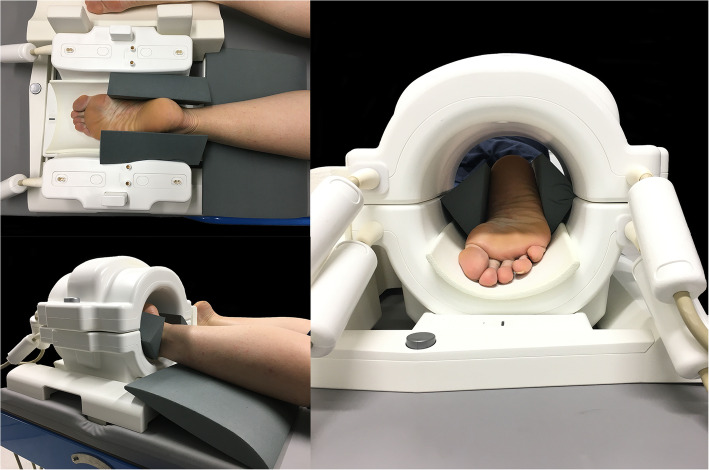
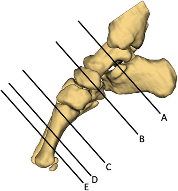
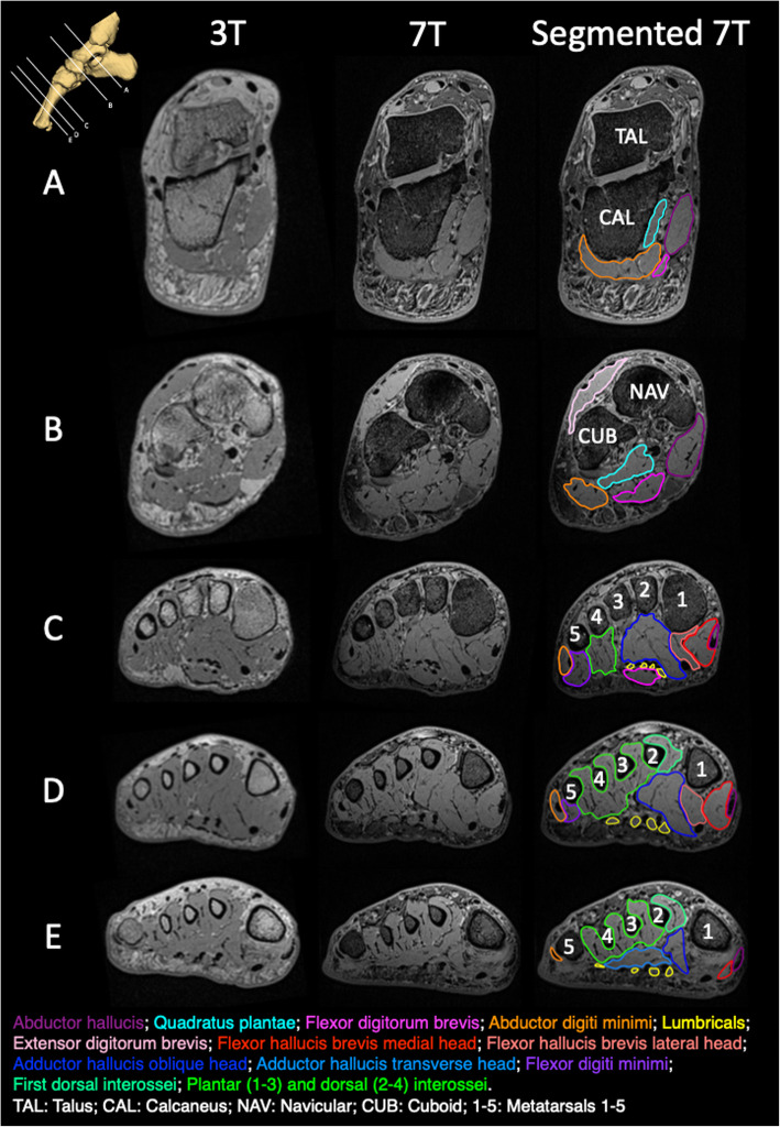
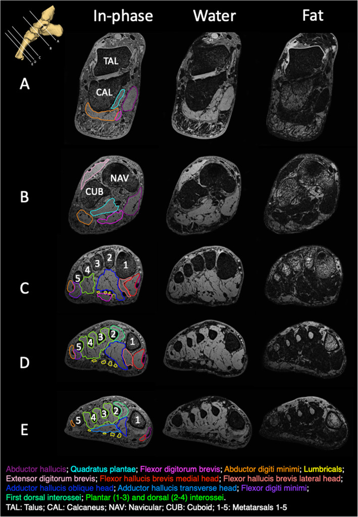
One healthy female (age 39 years, mass 65 kg, height 1.73 m) underwent MRI. A T1-weighted VIBE - radio-frequency spoiled 3D steady state GRE - sequence of the whole foot was acquired on a Siemens 7T MAGNETOM scanner, as well as a 3T MAGNETOM Prisma scanner for comparison. A high-resolution fat/water separation image was also acquired using a 3D 2-point DIXON sequence at 7T. Coronal plane images from 3T and 7T scanners were compared. Using 3D Slicer software, regions of interest were manually contoured for each muscle on 7T images. Muscle volumes and percentage of muscle fat infiltration were calculated (muscle fat infiltration % = Fat/(Fat + Water) x100) for each muscle.
Compared to the 3T images, the 7T images provided superior resolution, particularly at the forefoot, to facilitate segmentation of individual muscles. Muscle volumes ranged from 1.5 cm and 19.8 cm, and percentage muscle fat infiltration ranged from 9.2-15.0%.
This proof-of-concept study demonstrates a feasible method of quantifying muscle morphology and composition for individual intrinsic foot muscles using advanced high-field MRI techniques. This method can be used in future studies to better understand intrinsic foot muscle morphology and composition in healthy individuals, as well as those with lower disorders.
足部内在肌是足部功能的重要贡献者,在下肢疾病的评估中很重要。磁共振成像(MRI)提供了一种非侵入性的选择,可以测量肌肉形态和组成,这是肌肉功能的主要决定因素。超高场(7-T)磁共振成像提供了足够的信号来评估内在足部肌肉的形态,并且当与化学位移序列结合时,可以获得肌肉成分的测量值。在这里,我们旨在提供一种使用高场 MRI 测量内在足部肌肉形态和组成的概念验证方法。
一名健康女性(年龄 39 岁,体重 65 公斤,身高 1.73 米)接受了 MRI 检查。使用西门子 7T MAGNETOM 扫描仪采集了整个足部的 T1 加权 VIBE-射频扰相 3D 稳态 GRE 序列,以及用于比较的 3T MAGNETOM Prisma 扫描仪。还在 7T 上使用 3D 2 点 DIXON 序列采集了高分辨率的脂肪/水分离图像。比较了 3T 和 7T 扫描仪的冠状面图像。使用 3D Slicer 软件,手动在 7T 图像上为每个肌肉勾勒感兴趣区。计算了每个肌肉的肌肉体积和肌肉脂肪浸润百分比(肌肉脂肪浸润%=脂肪/(脂肪+水)×100)。
与 3T 图像相比,7T 图像提供了更好的分辨率,特别是在前足部,便于对各个肌肉进行分割。肌肉体积范围为 1.5cm 至 19.8cm,肌肉脂肪浸润百分比范围为 9.2-15.0%。
这项概念验证研究表明,使用先进的高场 MRI 技术量化个体内在足部肌肉的形态和组成是一种可行的方法。这种方法可用于未来的研究,以更好地了解健康个体以及下肢疾病患者的内在足部肌肉形态和组成。