• 文献检索
  • 文档翻译
  • 深度研究
  • 学术资讯
  • Suppr Zotero 插件Zotero 插件
  • 邀请有礼
  • 套餐&价格
  • 历史记录
应用&插件
Suppr Zotero 插件Zotero 插件浏览器插件Mac 客户端Windows 客户端微信小程序
定价
高级版会员购买积分包购买API积分包
服务
文献检索文档翻译深度研究API 文档MCP 服务
关于我们
关于 Suppr公司介绍联系我们用户协议隐私条款
关注我们

Suppr 超能文献

核心技术专利:CN118964589B侵权必究
粤ICP备2023148730 号-1Suppr @ 2026

文献检索

告别复杂PubMed语法,用中文像聊天一样搜索,搜遍4000万医学文献。AI智能推荐,让科研检索更轻松。

立即免费搜索

文件翻译

保留排版,准确专业,支持PDF/Word/PPT等文件格式,支持 12+语言互译。

免费翻译文档

深度研究

AI帮你快速写综述,25分钟生成高质量综述,智能提取关键信息,辅助科研写作。

立即免费体验

使用功能超声对啮齿动物的深部脑刺激反应进行成像。

Imaging the response to deep brain stimulation in rodent using functional ultrasound.

机构信息

Department of Radiology, Mayo Clinic College of Medicine and Science, Rochester, Minnesota 55902, United States.

Department of Neurologic Surgery, Mayo Clinic College of Medicine and Science, Rochester, Minnesota 55902, United States.

出版信息

Phys Med Biol. 2021 Feb 25;66(5):05LT01. doi: 10.1088/1361-6560/abdee5.

DOI:10.1088/1361-6560/abdee5
PMID:33482648
原文链接:https://pmc.ncbi.nlm.nih.gov/articles/PMC7920924/
Abstract

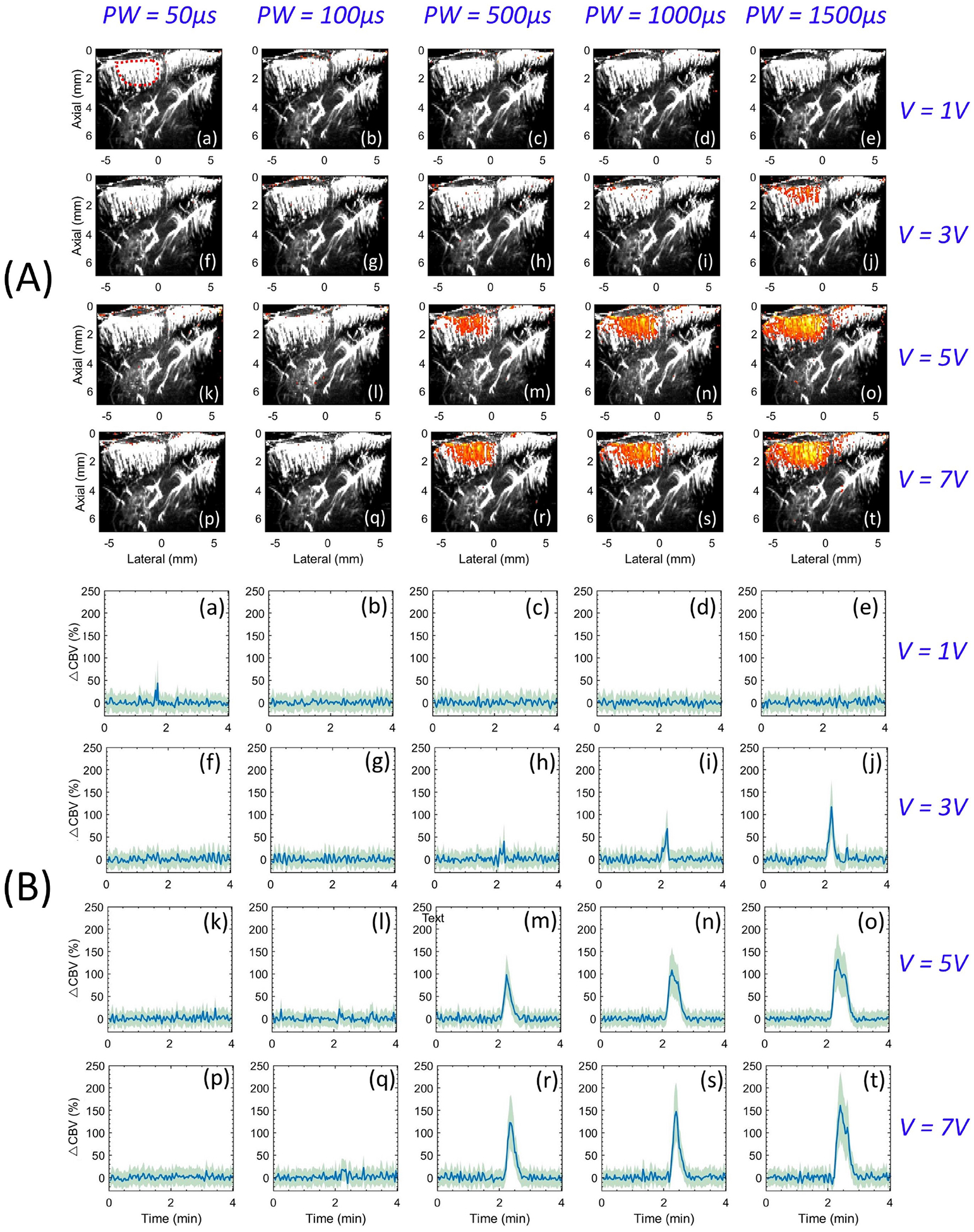
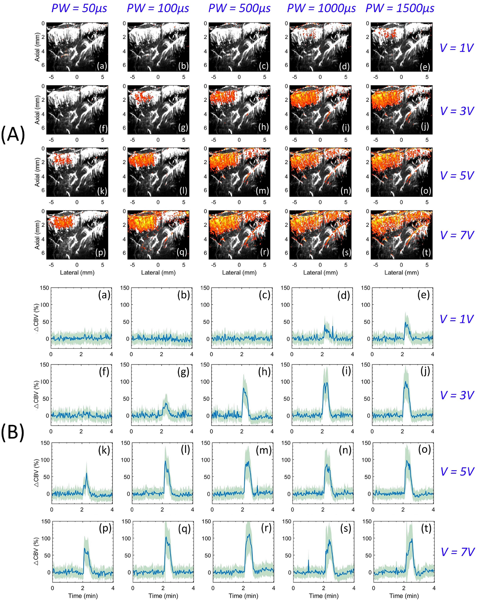
In this study, we explored the feasibility of using functional ultrasound (fUS) imaging to visualize cerebral activation associated with thalamic deep brain stimulation (DBS), in rodents. The ventrolateral (VL) thalamus was stimulated using electrical pulses of low and high frequencies of 10 and 100 Hz, respectively, and multiple voltages (1-7 V) and pulse widths (50-1500 μs). The fUS imaging demonstrated DBS-evoked activation of cerebral cortex based on changes of cerebral blood volume, specifically at the primary motor cortex (PMC). Low frequency stimulation (LFS) demonstrated significantly higher PMC activation compared to higher frequency stimulation (HFS), at intensities (5-7 V). Whereas, at lower intensities (1-3 V), only HFS demonstrated visible PMC activation. Further, LFS-evoked cerebral activation was was primarily located at the PMC. Our data presents the functionality and feasibility of fUS imaging as an investigational tool to identify brain areas associated with DBS. This preliminary study is an important stepping stone towards conducting real-time functional ultrasound imaging of DBS in awake and behaving animal models, which is of significant interest to the community for studying motor-related disorders.

摘要

在这项研究中,我们探索了使用功能超声(fUS)成像来可视化与丘脑深部脑刺激(DBS)相关的大脑激活的可行性,在啮齿动物中。使用低频和高频电脉冲分别为 10Hz 和 100Hz 刺激腹外侧(VL)丘脑,多个电压(1-7V)和脉冲宽度(50-1500μs)。fUS 成像基于脑血容量的变化显示了 DBS 诱发的大脑皮层激活,特别是在初级运动皮层(PMC)。与高频刺激(HFS)相比,低频刺激(LFS)在强度(5-7V)下表现出明显更高的 PMC 激活。然而,在较低强度(1-3V)下,只有 HFS 显示出可见的 PMC 激活。此外,LFS 诱发的大脑激活主要位于 PMC。我们的数据表明,fUS 成像作为一种研究工具具有功能性和可行性,可用于识别与 DBS 相关的大脑区域。这项初步研究是朝着在清醒和行为动物模型中进行实时功能超声成像 DBS 迈出的重要一步,这对于研究与运动相关的疾病的社区具有重要意义。

https://cdn.ncbi.nlm.nih.gov/pmc/blobs/979a/7920924/fd5cb5d15d14/nihms-1664690-f0005.jpg
https://cdn.ncbi.nlm.nih.gov/pmc/blobs/979a/7920924/7e033fb7c661/nihms-1664690-f0001.jpg
https://cdn.ncbi.nlm.nih.gov/pmc/blobs/979a/7920924/5be203511ffa/nihms-1664690-f0002.jpg
https://cdn.ncbi.nlm.nih.gov/pmc/blobs/979a/7920924/7c23ddc5b9ba/nihms-1664690-f0003.jpg
https://cdn.ncbi.nlm.nih.gov/pmc/blobs/979a/7920924/dc40e856b7b6/nihms-1664690-f0004.jpg
https://cdn.ncbi.nlm.nih.gov/pmc/blobs/979a/7920924/fd5cb5d15d14/nihms-1664690-f0005.jpg

相似文献

1
Imaging the response to deep brain stimulation in rodent using functional ultrasound.使用功能超声对啮齿动物的深部脑刺激反应进行成像。
Phys Med Biol. 2021 Feb 25;66(5):05LT01. doi: 10.1088/1361-6560/abdee5.
2
Frequency-dependent functional neuromodulatory effects on the motor network by ventral lateral thalamic deep brain stimulation in swine.腹外侧丘脑深部脑刺激对猪运动网络的频率依赖性功能神经调节作用。
Neuroimage. 2015 Jan 15;105:181-8. doi: 10.1016/j.neuroimage.2014.09.064. Epub 2014 Oct 14.
3
Spatiotemporal dynamics of cortical perfusion in response to thalamic deep brain stimulation.丘脑深部脑刺激响应下皮质灌注的时空动力学
Neuroimage. 2016 Feb 1;126:131-9. doi: 10.1016/j.neuroimage.2015.11.017. Epub 2015 Nov 11.
4
Frequency-dependent spike-pattern changes in motor cortex during thalamic deep brain stimulation.在丘脑深部脑刺激期间,运动皮层中依赖频率的尖峰模式变化。
J Neurophysiol. 2020 Nov 1;124(5):1518-1529. doi: 10.1152/jn.00198.2020. Epub 2020 Sep 23.
5
Orientation-selective and directional deep brain stimulation in swine assessed by functional MRI at 3T.3T 功能磁共振成像评估猪的取向选择性和方向选择性深部脑刺激。
Neuroimage. 2021 Jan 1;224:117357. doi: 10.1016/j.neuroimage.2020.117357. Epub 2020 Sep 9.
6
Changes in cortical excitability with thalamic deep brain stimulation.丘脑深部脑刺激引起的皮质兴奋性变化。
Neurology. 2005 Jun 14;64(11):1913-9. doi: 10.1212/01.WNL.0000163985.89444.DD.
7
Deep brain stimulation of the ventroanterior and ventrolateral thalamus improves motor function in a rat model of Parkinson's disease.腹侧前丘脑和腹外侧丘脑的深部脑刺激可改善帕金森病大鼠模型的运动功能。
Exp Neurol. 2019 Jul;317:155-167. doi: 10.1016/j.expneurol.2019.03.008. Epub 2019 Mar 16.
8
Neurovascular coupling during deep brain stimulation.深部脑刺激期间的神经血管耦联。
Brain Stimul. 2020 May-Jun;13(3):916-927. doi: 10.1016/j.brs.2020.03.005. Epub 2020 Mar 13.
9
Subthalamic deep brain stimulation alters neuronal firing in canonical pain nuclei in a 6-hydroxydopamine lesioned rat model of Parkinson's disease.丘脑底核深部脑刺激改变帕金森病 6-羟多巴胺损伤大鼠模型中经典痛核神经元的放电。
Exp Neurol. 2016 Sep;283(Pt A):298-307. doi: 10.1016/j.expneurol.2016.06.031. Epub 2016 Jul 1.
10
Model-based deconstruction of cortical evoked potentials generated by subthalamic nucleus deep brain stimulation.基于模型的丘脑底核深部脑刺激产生的皮质诱发电位解构
J Neurophysiol. 2018 Aug 1;120(2):662-680. doi: 10.1152/jn.00862.2017. Epub 2018 Apr 25.

引用本文的文献

1
Background Noise Removal in Non-Contrast- Enhanced Ultrasound Microvasculature Imaging Using Combined Collaborative, Morphological, and Vesselness Filtering.基于协同、形态学和血管性滤波相结合的非增强超声微血管成像背景噪声去除
IEEE Access. 2024;12:116150-116161. doi: 10.1109/access.2024.3446531. Epub 2024 Aug 20.
2
Theta-frequency medial septal nucleus deep brain stimulation increases neurovascular activity in MK-801-treated mice.θ波频率内侧隔核深部脑刺激可增加MK-801处理小鼠的神经血管活性。
Front Neurosci. 2024 Mar 15;18:1372315. doi: 10.3389/fnins.2024.1372315. eCollection 2024.
3
Mapping Pharmacologically Evoked Neurovascular Activation and Its Suppression in a Rat Model of Tremor Using Functional Ultrasound: A Feasibility Study.

本文引用的文献

1
Transfer functions linking neural calcium to single voxel functional ultrasound signal.将神经钙与单像素功能超声信号联系起来的传递函数。
Nat Commun. 2020 Jun 11;11(1):2954. doi: 10.1038/s41467-020-16774-9.
2
Adaptive background noise bias suppression in contrast-free ultrasound microvascular imaging.无对比超声微血管成像中的自适应背景噪声偏差抑制。
Phys Med Biol. 2019 Dec 19;64(24):245015. doi: 10.1088/1361-6560/ab5879.
3
4D Functional Imaging of the Rat Brain Using a Large Aperture Row-Column Array.使用大孔径行-列阵对大鼠大脑进行 4D 功能成像。
应用功能超声研究震颤大鼠模型中药物诱发的神经血管激活及其抑制的定位:一项可行性研究。
Sensors (Basel). 2023 Aug 3;23(15):6902. doi: 10.3390/s23156902.
4
[The developments and applications of functional ultrasound imaging].[功能超声成像的发展与应用]
Sheng Wu Yi Xue Gong Cheng Xue Za Zhi. 2022 Oct 25;39(5):1015-1021. doi: 10.7507/1001-5515.202206050.
IEEE Trans Med Imaging. 2020 Jun;39(6):1884-1893. doi: 10.1109/TMI.2019.2959833. Epub 2019 Dec 16.
4
Altered Primary Motor Cortex Neuronal Activity in a Rat Model of Harmaline-Induced Tremor During Thalamic Deep Brain Stimulation.在丘脑深部脑刺激期间,harmaline诱导的震颤大鼠模型中初级运动皮层神经元活动的改变
Front Cell Neurosci. 2019 Oct 15;13:448. doi: 10.3389/fncel.2019.00448. eCollection 2019.
5
4D functional ultrasound imaging of whole-brain activity in rodents.鼠脑全脑活动的 4D 功能超声成像。
Nat Methods. 2019 Oct;16(10):994-997. doi: 10.1038/s41592-019-0572-y. Epub 2019 Sep 23.
6
Functional ultrasound imaging of the brain reveals propagation of task-related brain activity in behaving primates.功能超声脑成像揭示了行为灵长类动物中与任务相关的脑活动的传播。
Nat Commun. 2019 Mar 28;10(1):1400. doi: 10.1038/s41467-019-09349-w.
7
Whole-Brain Functional Ultrasound Imaging Reveals Brain Modules for Visuomotor Integration.全脑功能超声成像揭示了视动整合的大脑模块。
Neuron. 2018 Dec 5;100(5):1241-1251.e7. doi: 10.1016/j.neuron.2018.11.031.
8
3D functional ultrasound imaging of pigeons.鸽子的 3D 功能超声成像。
Neuroimage. 2018 Dec;183:469-477. doi: 10.1016/j.neuroimage.2018.08.014. Epub 2018 Aug 14.
9
Multi-scale mapping along the auditory hierarchy using high-resolution functional UltraSound in the awake ferret.使用清醒雪貂的高分辨率功能超声在听觉层次上进行多尺度映射。
Elife. 2018 Jun 28;7:e35028. doi: 10.7554/eLife.35028.
10
Vascular Compartmentalization of Functional Hyperemia from the Synapse to the Pia.从突触到软脑膜的功能充血的血管分隔。
Neuron. 2018 Jul 25;99(2):362-375.e4. doi: 10.1016/j.neuron.2018.06.012. Epub 2018 Jun 21.