Rosshirt Nils, Trauth Richard, Platzer Hadrian, Tripel Elena, Nees Timo A, Lorenz Hanns-Martin, Tretter Theresa, Moradi Babak
Clinic for Orthopedic and Trauma Surgery, University Hospital Heidelberg, Schlierbacher Landstr. 200a, Heidelberg, 69118, Germany.
Department of Internal Medicine V, Division of Rheumatology, University Hospital Heidelberg, Heidelberg, Germany.
Arthritis Res Ther. 2021 Jan 22;23(1):37. doi: 10.1186/s13075-020-02410-w.
Investigating the pathophysiological mechanisms of early osteoarthritis (OA) is of utmost interest since this stage holds the strongest promise for therapeutic interventions. The aims of this study were to analyze if synovial inflammation is already present in early OA and to characterize the involved cell populations, by investigating synovial fluid (SF) and synovial membrane (SM) of early OA patients for the presence and polarization status of CD4 T cells.
A quantitative analysis of CD4 T cell infiltration in SF and SM compared to peripheral blood (PB) was performed in patients with early stages of OA. We further investigated intracellular staining (ICS), surface marker, and chemokine receptor expression profiles of CD4 T cells in SF, SM, and PB, as well as cytokine expression in native SF and PB. Matched samples of SF, SM, and PB were harvested from 40 patients with early OA at the time of surgery. Early OA was confirmed by independent surgeons intraoperatively. Samples were analyzed by flow cytometry for surface markers and cytokines, which are preferentially expressed by distinct T cell subsets (Th1, Th2, Th17, regulatory T cells). Furthermore, we analyzed native SF and PB supernatants using MACSPlex for multiple cytokine expression profiles.
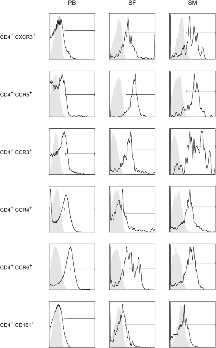
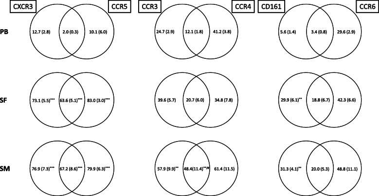
SF and SM showed a distinct infiltration of CD4 T lymphocytes, with significantly increased expression of chemokine receptors CXCR3/CCR5, cytokine IFN-γ (preferentially expressed by Th1 cells), and CD161 (preferentially expressed by IL-17 producing Th17 cells) compared to PB. Furthermore, the percentage of CD4 T cells polarized to Treg was significantly increased in SM compared to SF and PB. No significant differences were observed for CCR3 and CCR4 (preferentially expressed by Th2 cells), although IL-4 values were significantly higher in SM and SF compared to PB. Cytokine analysis showed comparable results between PB and SF, with only IL-6 being significantly increased in SF.
Early OA joints show already significant inflammation through CD4 T cell infiltration, with predominant Th1 cell polarization. Inflammation seems to be driven by direct proinflammatory cell interaction. Cytokine signaling seems to be negligible at the site of inflammation in early OA, with only IL-6 being significantly increased in SF compared to PB.
研究早期骨关节炎(OA)的病理生理机制至关重要,因为这一阶段对于治疗干预最具前景。本研究的目的是通过调查早期OA患者的滑液(SF)和滑膜(SM)中CD4 T细胞的存在情况和极化状态,分析早期OA中是否已经存在滑膜炎症,并对相关细胞群体进行特征描述。
对早期OA患者的SF和SM中CD4 T细胞浸润情况与外周血(PB)进行定量分析。我们进一步研究了SF、SM和PB中CD4 T细胞的细胞内染色(ICS)、表面标志物和趋化因子受体表达谱,以及天然SF和PB中的细胞因子表达。在手术时从40例早期OA患者中采集匹配的SF、SM和PB样本。早期OA由独立外科医生在术中确认。通过流式细胞术分析样本的表面标志物和细胞因子,这些标志物和细胞因子由不同的T细胞亚群(Th1、Th2、Th17、调节性T细胞)优先表达。此外,我们使用MACSPlex分析天然SF和PB上清液的多种细胞因子表达谱。
与PB相比,SF和SM显示出明显的CD4 T淋巴细胞浸润,趋化因子受体CXCR3/CCR5、细胞因子IFN-γ(由Th1细胞优先表达)和CD161(由产生IL-17的Th17细胞优先表达)的表达显著增加。此外,与SF和PB相比,SM中极化至Treg的CD4 T细胞百分比显著增加。CCR3和CCR4(由Th2细胞优先表达)未观察到显著差异,尽管与PB相比,SM和SF中的IL-4值显著更高。细胞因子分析显示PB和SF之间结果相当,只有SF中的IL-6显著增加。
早期OA关节通过CD4 T细胞浸润已经显示出明显的炎症,主要为Th1细胞极化。炎症似乎由直接的促炎细胞相互作用驱动。在早期OA的炎症部位,细胞因子信号似乎可以忽略不计,与PB相比,只有SF中的IL-6显著增加。