Voter Andrew F, Kanne Jeffrey P, Kuner Anthony D, Gettle Lori Mankowski
University of Wisconsin School of Medicine and Public Health, 600 Highland Avenue, Madison, WI 53705, USA.
Department of Radiology, University of Wisconsin School of Medicine and Public Health, 600 Highland Avenue, D4-352, Madison, WI 53705, USA.
Radiol Case Rep. 2021 Jan 12;16(3):704-706. doi: 10.1016/j.radcr.2020.12.066. eCollection 2021 Mar.
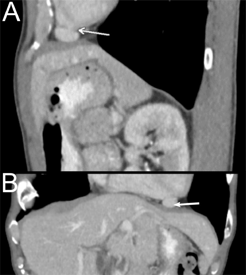
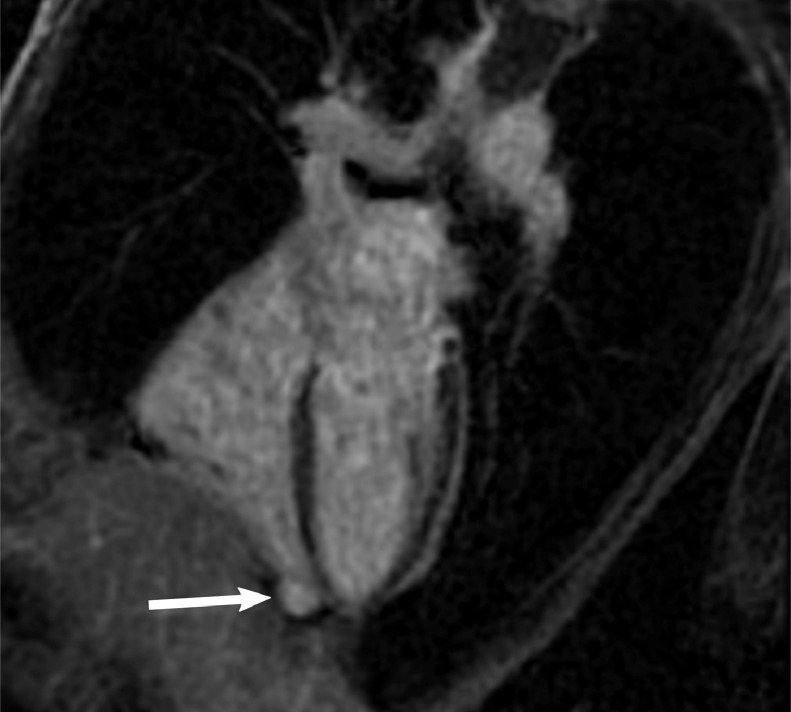
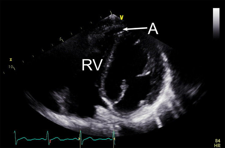
Three types of cardiac outpouchings are encountered on cardiovascular imaging: diverticula, aneurysms and pseudoaneurysms. The underlying physiology, imaging findings, risk of rupture, and optimal treatment varies for each and a correct diagnosis is critical. We report a case of a rare, incidentally discovered right ventricular aneurysm that was characterized by transthoracic echocardiogram, computed tomography, and cardiac MRI. The types of cardiac outpouchings are reviewed, and we discuss the selection of imaging modality, keys to distinguishing the outpouchings, and management strategies.
憩室、动脉瘤和假性动脉瘤。每种类型的潜在生理学、影像学表现、破裂风险及最佳治疗方法各不相同,正确诊断至关重要。我们报告一例罕见的、偶然发现的右心室动脉瘤病例,该病例通过经胸超声心动图、计算机断层扫描和心脏磁共振成像进行了特征性描述。本文回顾了心脏外凸的类型,并讨论了成像方式的选择、区分这些外凸的关键以及管理策略。