Department of Pathology and Cytology, Halland Hospital Halmstad, Halmstad, Sweden.
Division of Pathology, Department of Clinical Sciences Lund, Lund University, Lund, Sweden.
Cancer Cytopathol. 2021 Jun;129(6):468-478. doi: 10.1002/cncy.22401. Epub 2021 Jan 25.
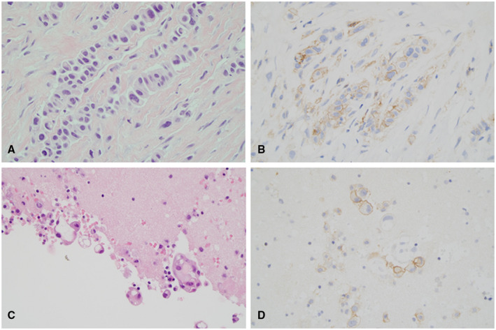
Malignant mesothelioma (MM) is a therapy-resistant tumor, often causing an effusion. Drugs targeting the programmed cell death 1 (PD-1)/programmed cell death ligand 1 (PD-L1) pathway have shown promising results, but assessment of PD-L1 expression to select patients for therapy has mainly been performed on histologic tissue samples. In a previous study, we showed that MM effusions are suitable for PD-L1 assessment with results comparable to those reported in histologic studies, but no studies have compared PD-L1 expression in histologic and cytologic samples.
PD-L1 expression was determined immunohistochemically (clone 28-8) in 61 paired samples of effusions and biopsies from patients with pleural MM, obtained at the time of diagnosis. Only cases with >100 tumor cells were included. Membranous staining in tumor cells was considered positive at ≥1%, >5%, >10%, and >50% cutoff levels.
Of 61 histologic samples, PD-L1 expression was found in 28 and 7 samples at ≥1% and >50% cutoffs, respectively; the corresponding figures for cytology were 21 and 5, respectively. The overall percentage agreement between histology and cytology was 69% and 84%, with a kappa (κ) of 0.36 and 0.08 at ≥1% and >50% cutoffs, respectively. The concordance between cytology and histology tended to be higher for epithelioid MM versus nonepithelioid MM at a ≥1% cutoff. PD-L1 positivity in biopsies, but not in effusions, correlated with the histologic subtype at a ≥1% cutoff.
A moderate concordance of PD-L1 expression between biopsies and effusions from pleural MM, especially for the epithelioid subtype, indicates biological differences between the 2 types of specimens. Cytology and histology may be complementary.
恶性间皮瘤(MM)是一种治疗耐药的肿瘤,常导致胸腔积液。针对程序性细胞死亡 1(PD-1)/程序性细胞死亡配体 1(PD-L1)通路的药物已显示出良好的效果,但评估 PD-L1 表达以选择治疗患者主要是在组织学组织样本上进行的。在之前的一项研究中,我们表明 MM 胸腔积液适合进行 PD-L1 评估,结果与组织学研究报告的结果相当,但尚无研究比较组织学和细胞学样本中的 PD-L1 表达。
在诊断时,对 61 例胸膜 MM 患者的胸腔积液和活检样本进行免疫组织化学(克隆 28-8)检测 PD-L1 表达。仅纳入有>100 个肿瘤细胞的病例。肿瘤细胞的膜状染色被认为在≥1%、>5%、>10%和>50%截断值时为阳性。
在 61 例组织学样本中,分别有 28 例和 7 例在≥1%和>50%截断值时检测到 PD-L1 表达;细胞学样本分别为 21 例和 5 例。组织学和细胞学之间的总体百分比一致性分别为 69%和 84%,在≥1%和>50%截断值时,κ值分别为 0.36 和 0.08。在≥1%截断值时,上皮样 MM 与非上皮样 MM 之间,细胞学与组织学的一致性较高。活检中 PD-L1 的阳性率,但不是胸腔积液中的阳性率,与≥1%截断值时的组织学亚型相关。
胸膜 MM 活检和胸腔积液中 PD-L1 表达之间存在中度一致性,尤其是上皮样亚型,表明这两种标本之间存在生物学差异。细胞学和组织学可能是互补的。