CERVO Brain Research Centre, Quebec, QC, Canada.
Department of Physiology and Cell Biology, Ohio State University, Columbus, OH, USA.
Sci Rep. 2021 Jan 28;11(1):2500. doi: 10.1038/s41598-021-82007-8.
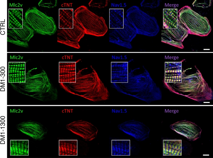
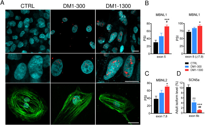
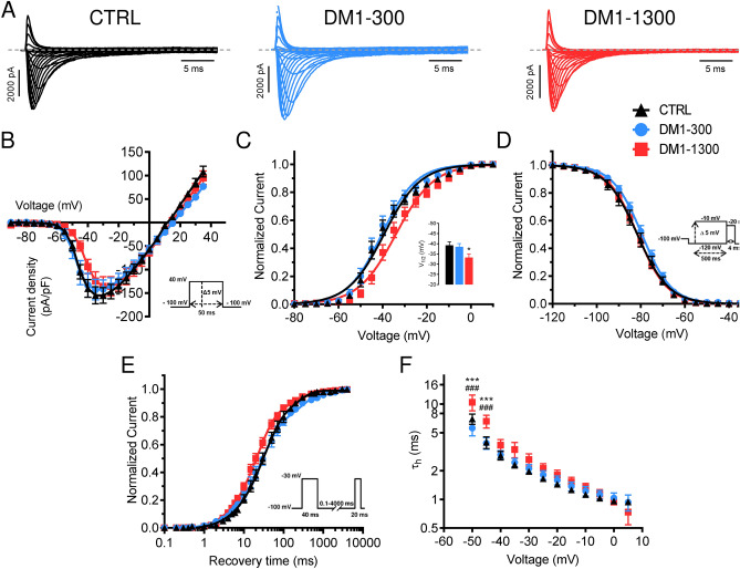
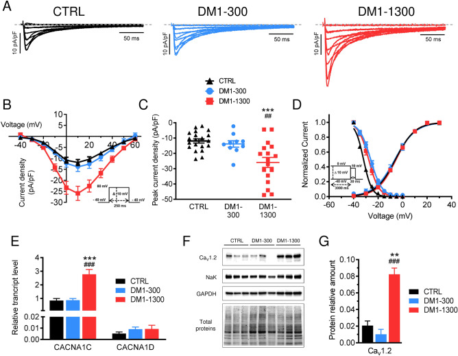
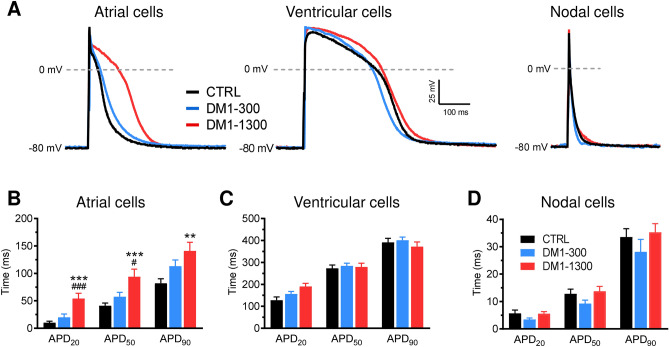
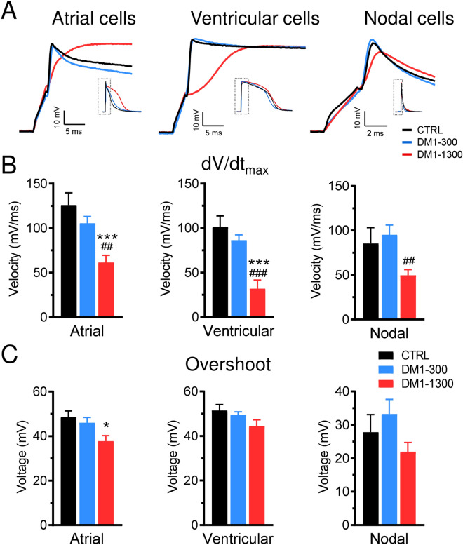
Cardiac complications such as electrical abnormalities including conduction delays and arrhythmias are the main cause of death in individuals with Myotonic Dystrophy type 1 (DM1). We developed a disease model using iPSC-derived cardiomyocytes (iPSC-CMs) from a healthy individual and two DM1 patients with different CTG repeats lengths and clinical history (DM1-1300 and DM1-300). We confirmed the presence of toxic RNA foci and mis-spliced MBNL1/2 transcripts in DM1 iPSC-CMs. In DM1-1300, we identified a switch in the cardiac sodium channel SCN5A from the adult to the neonatal isoform. The down-regulation of adult SCN5A isoforms is consistent with a shift in the sodium current activation to depolarized potentials observed in DM1-1300. L-type calcium current density was higher in iPSC-CMs from DM1-1300, which is correlated with the overexpression of the Ca1.2 transcript and proteins. Importantly, I and I dysfunctions resulted in prolonged action potentials duration, slower velocities, and decreased overshoots. Optical mapping analysis revealed a slower conduction velocity in DM1-1300 iPSC-CM monolayers. In conclusion, our data revealed two distinct ions channels perturbations in DM1 iPSC-CM from the patient with cardiac dysfunction, one affecting Na channels and one affecting Ca channels. Both have an impact on cardiac APs and ultimately on heart conduction.
心脏并发症,如传导延迟和心律失常等电异常,是 1 型肌强直性营养不良(DM1)患者死亡的主要原因。我们使用来自健康个体和两名具有不同 CTG 重复长度和临床病史的 DM1 患者的 iPSC 衍生心肌细胞(iPSC-CM)开发了一种疾病模型(DM1-1300 和 DM1-300)。我们证实了 DM1 iPSC-CM 中存在毒性 RNA 焦点和错误拼接的 MBNL1/2 转录本。在 DM1-1300 中,我们发现心脏钠离子通道 SCN5A 从成人型转变为新生儿型。成人 SCN5A 同工型的下调与在 DM1-1300 中观察到的钠离子电流激活向去极化电位的转变一致。DM1-1300 中的 iPSC-CM 中的 L 型钙电流密度更高,这与 Ca1.2 转录本和蛋白的过度表达相关。重要的是,I 和 I 功能障碍导致动作电位持续时间延长、速度减慢和超射减少。光学映射分析显示 DM1-1300 iPSC-CM 单层中的传导速度较慢。总之,我们的数据揭示了心脏功能障碍患者的 DM1 iPSC-CM 中存在两种不同的离子通道扰动,一种影响 Na 通道,另一种影响 Ca 通道。这两者都对心脏 APs 产生影响,并最终对心脏传导产生影响。