Department of Nephrology, The Second Xiangya Hospital, Central South University, 139 Renmin Road, Changsha, 410011, Hunan, China.
Hunan Key Laboratory of Kidney Disease and Blood Purification, 139 Renmin Road, Changsha, 410011, Hunan, China.
BMC Nephrol. 2021 Jan 29;22(1):43. doi: 10.1186/s12882-021-02237-w.
Immunoglobulin A nephropathy (IgAN) is identified as mesangial IgA deposition and is usually accompanied by other immunofluorescence deposits. The impact of immunofluorescent features in IgAN patients, however, remains unclear.
Baseline clinicopathologic parameters and renal outcomes of 337 patients diagnosed with IgAN between January 2009 and December 2015 were analyzed. We then categorized these patients into four groups: without immunofluorescence deposits, mesangial-only, mesangial and glomerular capillary loops (GCLs), and GCLs-only. The study endpoint was end-stage kidney disease (ESKD) or a ≥ 50% decline in the estimated glomerular filtration rate (eGFR). Kaplan-Meier and Cox regression analyses were performed to calculate renal survival.
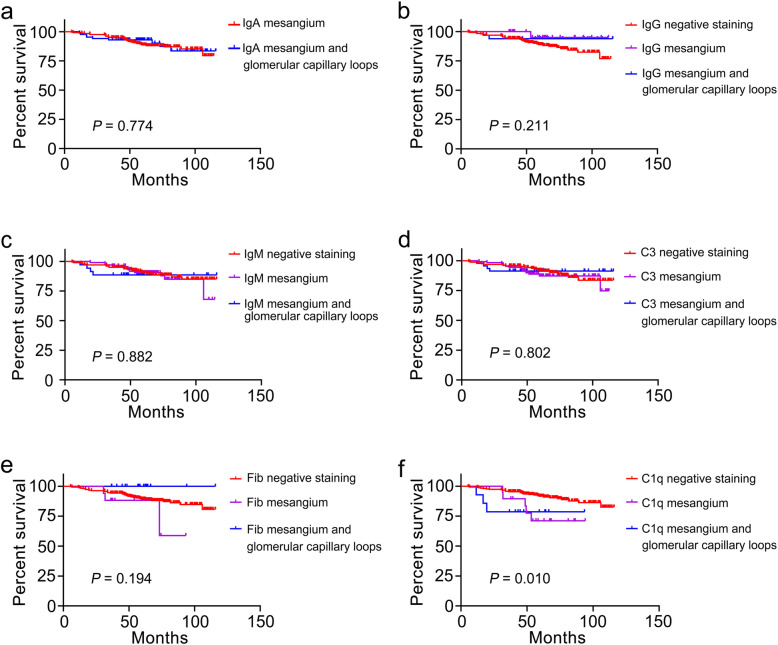
Of the 337 IgAN patients, women comprised 57.0%. Compared to patients with IgA deposition in the mesangial-only group, patients with IgA deposition in the mesangial +GCLs group were much heavier, and exhibited higher systolic blood pressure, lower serum IgG levels, and heavier proteinuria (all P < 0.05). Patients with IgG deposition in the mesangial +GCLs group presented with higher levels of cholesterol, heavier proteinuria than IgG deposition in the mesangial-only group (both P < 0.05). Compared with the mesangial-only group exhibiting C3 deposits, patients in the mesangial +GCLs group with C3 deposition had a higher systolic blood pressure (P = 0.028). A total of 38 patients (11.3%) continued to the study endpoint after a median follow-up time of 63.5 months (range,49.8-81.4). Kaplan-Meier analysis and Cox regression analysis showed that C1q deposition in the mesangial +GCLs group predicted a poor renal prognosis.
IgA and IgG deposits in the mesangial region and GCLs were associated with more unfavorable clinical and histopathologic findings in IgAN patients. C1q deposition in the mesangial region and GCLs predicted a poor renal prognosis. However, the impact of the pattern of immunofluorescence deposits on renal outcomes remains to be proven by further investigation.
免疫球蛋白 A 肾病(IgAN)的特征为系膜区 IgA 沉积,通常伴有其他免疫荧光沉积。然而,IgAN 患者免疫荧光特征的影响尚不清楚。
分析了 2009 年 1 月至 2015 年 12 月期间确诊的 337 例 IgAN 患者的基线临床病理参数和肾脏结局。然后,我们将这些患者分为四组:无免疫荧光沉积组、系膜区仅沉积组、系膜区和肾小球毛细血管袢(GCL)均沉积组和 GCL 仅沉积组。研究终点为终末期肾病(ESKD)或估算肾小球滤过率(eGFR)下降≥50%。采用 Kaplan-Meier 和 Cox 回归分析计算肾脏存活率。
337 例 IgAN 患者中,女性占 57.0%。与系膜区仅沉积组相比,系膜区+GCL 沉积组患者的体重明显增加,收缩压较高,血清 IgG 水平较低,蛋白尿较重(均 P<0.05)。系膜区+GCL 沉积组 IgG 沉积患者的胆固醇水平和蛋白尿均高于系膜区仅沉积组 IgG 沉积患者(均 P<0.05)。与系膜区仅沉积组 C3 沉积相比,系膜区+GCL 沉积组 C3 沉积患者的收缩压更高(P=0.028)。中位随访时间为 63.5 个月(范围 49.8-81.4)后,共有 38 例(11.3%)患者达到研究终点。Kaplan-Meier 分析和 Cox 回归分析显示,系膜区+GCL 沉积的 C1q 沉积预测肾脏预后不良。
IgAN 患者系膜区和 GCL 区的 IgA 和 IgG 沉积与更不利的临床和组织病理学发现相关。系膜区和 GCL 区的 C1q 沉积预测肾脏预后不良。然而,免疫荧光沉积模式对肾脏结局的影响仍需进一步研究证实。