Clinical Imaging Sciences Centre, Brighton and Sussex Medical School, Brighton, United Kingdom; Brighton and Sussex University Hospital Trust, Brighton, United Kingdom.
Neuroimaging Laboratory, Santa Lucia Foundation, Rome, Italy.
Neuroimage Clin. 2021;29:102562. doi: 10.1016/j.nicl.2021.102562. Epub 2021 Jan 14.
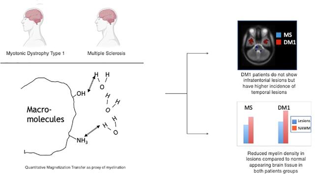
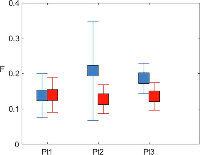
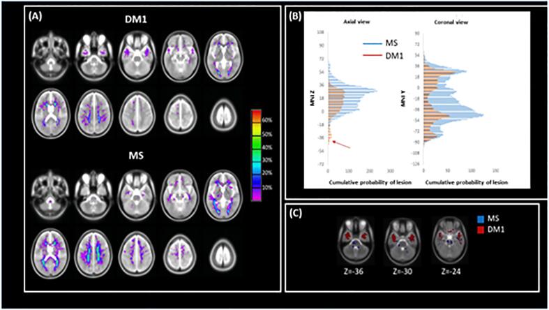
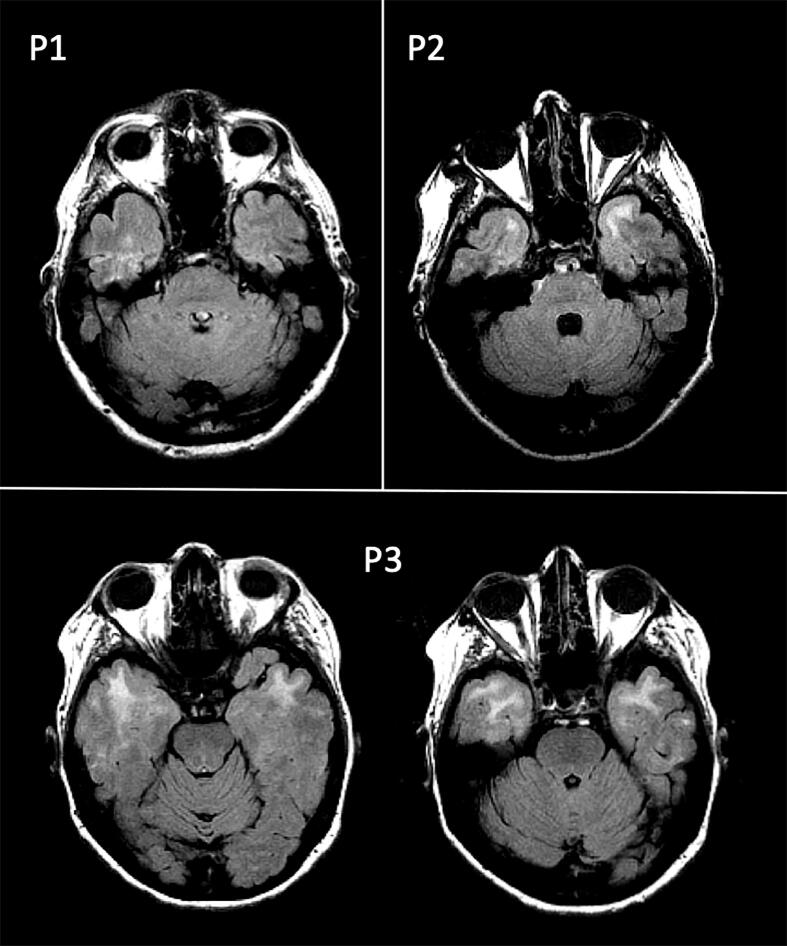
Myotonic Dystrophy type 1 (DM1) is an autosomal dominant condition caused by expansion of the CTG triplet repeats within the myotonic dystrophy protein of the kinase (DMPK) gene. The central nervous system is involved in the disease, with multiple symptoms including cognitive impairment. A typical feature of DM1 is the presence of widespread white matter (WM) lesions, whose total volume is associated with CTG triplet expansion. The aim of this study was to characterize the distribution and pathological substrate of these lesions as well as the normal appearing WM (NAWM) using quantitative magnetization transfer (qMT) MRI, and comparing data from DM1 patients with those from patients with multiple sclerosis (MS). Twenty-eight patients with DM1, 29 patients with relapsing-remitting MS, and 15 healthy controls had an MRI scan, including conventional and qMT imaging. The average pool size ratio (F), a proxy of myelination, was computed within lesions and NAWM for every participant. The lesion masks were warped into MNI space and lesion probability maps were obtained for each patient group. The lesion distribution, total lesion load and the tissue-specific mean F were compared between groups. The supratentorial distribution of lesions was similar in the 2 patient groups, although mean lesion volume was higher in MS than DM1. DM1 presented higher prevalence of anterior temporal lobe lesions, but none in the cerebellum and brainstem. Significantly reduced F values were found within DM1 lesions, suggesting a loss of myelin density. While F was reduced in the NAWM of MS patients, it did not differ between DM1 and controls. Our results provide further evidence for a need to compare histology and imaging using new MRI techniques in DM1 patients, in order to further our understanding of the underlying disease process contributing to WM disease.
1 型肌强直性营养不良(DM1)是一种常染色体显性疾病,由肌强直性营养不良蛋白激酶(DMPK)基因中的 CTG 三核苷酸重复扩展引起。中枢神经系统受累,多种症状包括认知障碍。DM1 的一个典型特征是存在广泛的白质(WM)病变,其总体积与 CTG 三核苷酸扩展有关。本研究的目的是使用定量磁化传递(qMT)MRI 来描述这些病变以及正常表现的 WM(NAWM)的分布和病理学基础,并将 DM1 患者的数据与多发性硬化症(MS)患者的数据进行比较。28 例 DM1 患者、29 例复发缓解型 MS 患者和 15 名健康对照者进行了 MRI 扫描,包括常规和 qMT 成像。为每位参与者计算了病变和 NAWM 内的平均池大小比(F),这是髓鞘化的一个代理指标。将病变掩模变形到 MNI 空间,并为每个患者组获得病变概率图。比较了各组之间的病变分布、总病变负荷和组织特异性平均 F。虽然 MS 患者的平均病变体积大于 DM1,但 2 组患者的病变上脑分布相似。DM1 患者更易出现前颞叶病变,但在小脑和脑干中均无病变。在 DM1 病变内发现 F 值显著降低,表明髓鞘密度丧失。虽然 MS 患者的 NAWM 中 F 值降低,但与 DM1 和对照组无差异。我们的研究结果进一步证明,需要使用新的 MRI 技术在 DM1 患者中比较组织学和影像学,以进一步了解导致 WM 疾病的潜在疾病过程。