• 文献检索
  • 文档翻译
  • 深度研究
  • 学术资讯
  • Suppr Zotero 插件Zotero 插件
  • 邀请有礼
  • 套餐&价格
  • 历史记录
应用&插件
Suppr Zotero 插件Zotero 插件浏览器插件Mac 客户端Windows 客户端微信小程序
定价
高级版会员购买积分包购买API积分包
服务
文献检索文档翻译深度研究API 文档MCP 服务
关于我们
关于 Suppr公司介绍联系我们用户协议隐私条款
关注我们

Suppr 超能文献

核心技术专利:CN118964589B侵权必究
粤ICP备2023148730 号-1Suppr @ 2026

文献检索

告别复杂PubMed语法,用中文像聊天一样搜索,搜遍4000万医学文献。AI智能推荐,让科研检索更轻松。

立即免费搜索

文件翻译

保留排版,准确专业,支持PDF/Word/PPT等文件格式,支持 12+语言互译。

免费翻译文档

深度研究

AI帮你快速写综述,25分钟生成高质量综述,智能提取关键信息,辅助科研写作。

立即免费体验

脑回状颅内出血和脑回状皮层:脑淀粉样血管病的新型磁共振成像标志物。

Striped occipital cortex and intragyral hemorrhage: Novel magnetic resonance imaging markers for cerebral amyloid angiopathy.

机构信息

Department of Neurology, 4501Leiden University Medical Center, Leiden, the Netherlands.

Department of Neurology and Neurosurgery, University Medical Center Utrecht Brain Center, Utrecht University, Utrecht, the Netherlands.

出版信息

Int J Stroke. 2021 Dec;16(9):1031-1038. doi: 10.1177/1747493021991961. Epub 2021 Feb 3.

DOI:10.1177/1747493021991961
PMID:33535905
原文链接:https://pmc.ncbi.nlm.nih.gov/articles/PMC8669214/
Abstract

BACKGROUND AND AIM

To investigate whether a striped occipital cortex and intragyral hemorrhage, two markers recently detected on ultra-high-field 7-tesla-magnetic resonance imaging in hereditary cerebral amyloid angiopathy (CAA), also occur in sporadic CAA (sCAA) or non-sCAA intracerebral hemorrhage (ICH).

METHODS

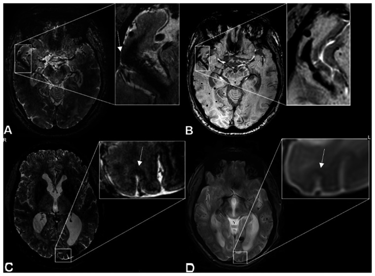
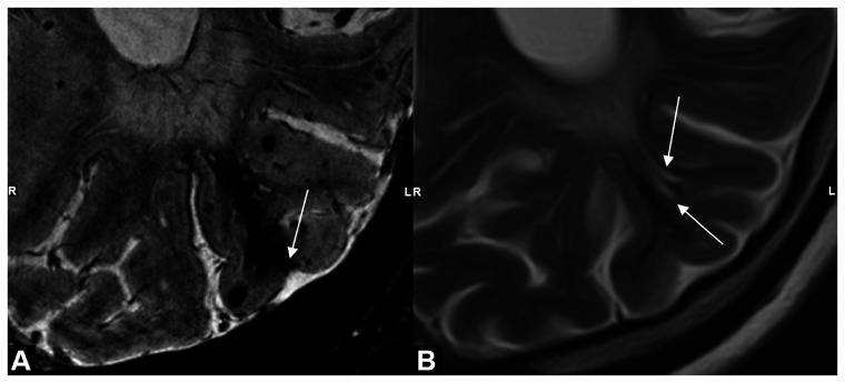
We performed 7-tesla-magnetic resonance imaging in patients with probable sCAA and patients with non-sCAA-ICH. Striped occipital cortex (linear hypointense stripes perpendicular to the cortex) and intragyral hemorrhage (hemorrhage restricted to the juxtacortical white matter of one gyrus) were scored on T*-weighted magnetic resonance imaging. We assessed the association between the markers, other CAA-magnetic resonance imaging markers and clinical features.

RESULTS

We included 33 patients with sCAA (median age 70 years) and 29 patients with non-sCAA-ICH (median age 58 years). Striped occipital cortex was detected in one (3%) patient with severe sCAA. Five intragyral hemorrhages were found in four (12%) sCAA patients. The markers were absent in the non-sCAA-ICH group. Patients with intragyral hemorrhages had more lobar ICHs (median count 6.5 vs. 1.0), lobar microbleeds (median count >50 vs. 15), and lower median cognitive scores (Mini Mental State Exam: 20 vs. 28, Montreal Cognitive Assessment: 18 vs. 24) compared with patients with sCAA without intragyral hemorrhage. In 12 (36%) patients, sCAA diagnosis was changed to mixed-type small vessel disease due to deep bleeds previously unobserved on lower field-magnetic resonance imaging.

CONCLUSION

Whereas a striped occipital cortex is rare in sCAA, 12% of patients with sCAA have intragyral hemorrhages. Intragyral hemorrhages seem to be related to advanced disease and their absence in patients with non-sCAA-ICH could suggest specificity for CAA.

摘要

背景与目的

本研究旨在探究条纹状枕叶皮质和脑内血肿(intragyral hemorrhage)两种在遗传性脑淀粉样血管病(hereditary cerebral amyloid angiopathy,CAA)超高清场 7 特斯拉磁共振成像(magnetic resonance imaging,MRI)中发现的标志物是否也存在于散发性 CAA(sporadic CAA,sCAA)或非 sCAA 脑出血(intracerebral hemorrhage,ICH)中。

方法

我们对疑似 sCAA 患者和非 sCAA-ICH 患者进行了 7 特斯拉 MRI 检查。在 T*-加权 MRI 上对条纹状枕叶皮质(垂直于皮质的线性低信号条纹)和脑内血肿(局限于一个脑回皮质下白质的血肿)进行评分。我们评估了这些标志物与其他 CAA-MRI 标志物和临床特征之间的关系。

结果

我们纳入了 33 例 sCAA 患者(中位年龄 70 岁)和 29 例非 sCAA-ICH 患者(中位年龄 58 岁)。1 例严重 sCAA 患者中发现 1 个条纹状枕叶皮质。4 例 sCAA 患者中发现 5 个脑内血肿。非 sCAA-ICH 组中未发现这些标志物。脑内血肿患者的脑叶 ICH 更多(中位数 6.5 个 vs. 1.0 个)、脑叶微出血(中位数>50 个 vs. 15 个)和更低的认知评分中位数(简易精神状态检查:20 分 vs. 28 分,蒙特利尔认知评估:18 分 vs. 24 分),与无脑内血肿的 sCAA 患者相比。由于先前在较低场 MRI 上未观察到深部出血,12 例(36%)患者的 sCAA 诊断更改为混合性小血管疾病。

结论

尽管条纹状枕叶皮质在 sCAA 中较为罕见,但 12%的 sCAA 患者存在脑内血肿。脑内血肿似乎与疾病的晚期有关,而非 sCAA-ICH 患者中无脑内血肿的存在可能提示其对 CAA 的特异性。

https://cdn.ncbi.nlm.nih.gov/pmc/blobs/ddfe/8669214/7fc4c698ff5c/10.1177_1747493021991961-fig2.jpg
https://cdn.ncbi.nlm.nih.gov/pmc/blobs/ddfe/8669214/372bde5982ec/10.1177_1747493021991961-fig1.jpg
https://cdn.ncbi.nlm.nih.gov/pmc/blobs/ddfe/8669214/7fc4c698ff5c/10.1177_1747493021991961-fig2.jpg
https://cdn.ncbi.nlm.nih.gov/pmc/blobs/ddfe/8669214/372bde5982ec/10.1177_1747493021991961-fig1.jpg
https://cdn.ncbi.nlm.nih.gov/pmc/blobs/ddfe/8669214/7fc4c698ff5c/10.1177_1747493021991961-fig2.jpg

相似文献

1
Striped occipital cortex and intragyral hemorrhage: Novel magnetic resonance imaging markers for cerebral amyloid angiopathy.脑回状颅内出血和脑回状皮层:脑淀粉样血管病的新型磁共振成像标志物。
Int J Stroke. 2021 Dec;16(9):1031-1038. doi: 10.1177/1747493021991961. Epub 2021 Feb 3.
2
Occipital Cortical Calcifications in Cerebral Amyloid Angiopathy.大脑淀粉样血管病中的枕叶皮质钙化。
Stroke. 2021 May;52(5):1851-1855. doi: 10.1161/STROKEAHA.120.033286. Epub 2021 Apr 5.
3
Spatial and temporal intracerebral hemorrhage patterns in Dutch-type hereditary cerebral amyloid angiopathy.荷兰型遗传性脑动脉淀粉样血管病的颅内出血时空模式。
Int J Stroke. 2022 Aug;17(7):793-798. doi: 10.1177/17474930211057022. Epub 2021 Nov 18.
4
Cerebral Amyloid Angiopathy With Vascular Iron Accumulation and Calcification.脑淀粉样血管病伴血管铁蓄积和钙化。
Stroke. 2018 Sep;49(9):2081-2087. doi: 10.1161/STROKEAHA.118.021872.
5
Neuropsychiatric symptoms with focus on apathy and irritability in sporadic and hereditary cerebral amyloid angiopathy.散发性和遗传性脑淀粉样血管病的神经精神症状,以冷漠和易怒为重点。
Alzheimers Res Ther. 2024 Apr 6;16(1):74. doi: 10.1186/s13195-024-01445-4.
6
Cerebellar Superficial Siderosis in Cerebral Amyloid Angiopathy.脑淀粉样血管病中的小脑表浅铁沉积症。
Stroke. 2022 Feb;53(2):552-557. doi: 10.1161/STROKEAHA.121.035019. Epub 2021 Sep 20.
7
Sex Differences in Onset and Progression of Cerebral Amyloid Angiopathy.性别差异与脑淀粉样血管病的发病和进展。
Stroke. 2023 Feb;54(2):306-314. doi: 10.1161/STROKEAHA.122.040823. Epub 2023 Jan 23.
8
Distribution of Lacunar Infarcts in Asians With Intracerebral Hemorrhage: A Magnetic Resonance Imaging and Amyloid Positron Emission Tomography Study.腔隙性脑梗死在亚洲人群中的分布:一项磁共振成像和淀粉样 PET 研究。
Stroke. 2018 Jun;49(6):1515-1517. doi: 10.1161/STROKEAHA.118.021539. Epub 2018 Apr 25.
9
Subarachnoid and Subdural Hemorrhages in Lobar Intracerebral Hemorrhage Associated With Cerebral Amyloid Angiopathy.脑叶脑出血合并脑淀粉样血管病相关的蛛网膜下腔出血和硬脑膜下出血。
Stroke. 2019 Jun;50(6):1567-1569. doi: 10.1161/STROKEAHA.119.024837. Epub 2019 Apr 16.
10
Cerebellar hemorrhages in patients with Dutch-type hereditary cerebral amyloid angiopathy.荷兰型遗传性脑动脉淀粉样血管病患者的小脑出血。
Int J Stroke. 2022 Jul;17(6):637-644. doi: 10.1177/17474930211043663. Epub 2021 Sep 10.

引用本文的文献

1
Updated imaging markers in cerebral amyloid angiopathy: What radiologists need to know.脑淀粉样血管病的更新影像学标志物:放射科医生需要了解的内容。
Jpn J Radiol. 2025 May;43(5):736-751. doi: 10.1007/s11604-024-01720-2. Epub 2024 Dec 28.
2
One Size Does Not Fit All: Micro-, Meso-, and Macrobleeds in Cerebral Amyloid Angiopathy.一刀切并不适用于所有情况:脑淀粉样血管病中的微出血、中出血和大出血
Cerebrovasc Dis. 2025;54(4):452-459. doi: 10.1159/000540899. Epub 2024 Aug 19.
3
Evaluating the role of 7-Tesla magnetic resonance imaging in neurosurgery: Trends in literature since clinical approval.

本文引用的文献

1
Cerebral Small Vessel Disease (CSVD) - Lessons From the Animal Models.脑小血管病(CSVD)——来自动物模型的经验教训。
Front Physiol. 2019 Oct 24;10:1317. doi: 10.3389/fphys.2019.01317. eCollection 2019.
2
Invited Review: The spectrum of age-related small vessel diseases: potential overlap and interactions of amyloid and nonamyloid vasculopathies.特邀综述:与年龄相关的小血管疾病谱:淀粉样和非淀粉样血管病的潜在重叠和相互作用。
Neuropathol Appl Neurobiol. 2020 Apr;46(3):219-239. doi: 10.1111/nan.12576. Epub 2019 Sep 11.
3
Cerebral Amyloid Angiopathy With Vascular Iron Accumulation and Calcification.
评估7特斯拉磁共振成像在神经外科手术中的作用:自临床获批以来的文献趋势。
World J Radiol. 2024 Jul 28;16(7):274-293. doi: 10.4329/wjr.v16.i7.274.
4
Occult Amyloid-β-Related Angiitis: Neuroimaging Findings at 1.5T, 3T, and 7T MRI.隐匿性β淀粉样蛋白相关血管炎:1.5T、3T 和 7T MRI 的神经影像学表现。
AJNR Am J Neuroradiol. 2024 Aug 9;45(8):1013-1018. doi: 10.3174/ajnr.A8264.
5
Histopathological Correlates of Lobar Microbleeds in False-Positive Cerebral Amyloid Angiopathy Cases.脑淀粉样血管病假阳性病例中微出血灶的组织病理学相关性
Ann Neurol. 2023 Nov;94(5):856-870. doi: 10.1002/ana.26761. Epub 2023 Sep 6.
脑淀粉样血管病伴血管铁蓄积和钙化。
Stroke. 2018 Sep;49(9):2081-2087. doi: 10.1161/STROKEAHA.118.021872.
4
Innovative Magnetic Resonance Imaging Markers of Hereditary Cerebral Amyloid Angiopathy at 7 Tesla.7 特斯拉下遗传性脑淀粉样血管病的创新性磁共振成像标志物。
Stroke. 2018 Jun;49(6):1518-1520. doi: 10.1161/STROKEAHA.117.020302. Epub 2018 Apr 25.
5
The Edinburgh CT and genetic diagnostic criteria for lobar intracerebral haemorrhage associated with cerebral amyloid angiopathy: model development and diagnostic test accuracy study.爱丁堡 CT 和与脑淀粉样血管病相关的脑叶颅内出血的基因诊断标准:模型建立和诊断试验准确性研究。
Lancet Neurol. 2018 Mar;17(3):232-240. doi: 10.1016/S1474-4422(18)30006-1. Epub 2018 Jan 10.
6
Mixed-location cerebral hemorrhage/microbleeds: Underlying microangiopathy and recurrence risk.混合部位脑出血/微出血:潜在的微血管病和复发风险。
Neurology. 2018 Jan 9;90(2):e119-e126. doi: 10.1212/WNL.0000000000004797. Epub 2017 Dec 15.
7
Cortical localization of microbleeds in cerebral amyloid angiopathy: an ultra high-field 7T MRI study.脑淀粉样血管病中微出血的皮质定位:一项超高场7T磁共振成像研究
J Alzheimers Dis. 2015;43(4):1325-30. doi: 10.3233/JAD-140864.
8
Neuroimaging standards for research into small vessel disease and its contribution to ageing and neurodegeneration.神经影像学标准研究小血管疾病及其对衰老和神经退行性变的影响。
Lancet Neurol. 2013 Aug;12(8):822-38. doi: 10.1016/S1474-4422(13)70124-8.
9
Cerebral amyloid angiopathy in the elderly.老年人脑淀粉样血管病。
Ann Neurol. 2011 Dec;70(6):871-80. doi: 10.1002/ana.22516.
10
7 tesla MRI of microbleeds and white matter lesions as seen in vascular dementia.7 特斯拉 MRI 对血管性痴呆患者微出血和白质病变的观察。
J Magn Reson Imaging. 2011 Apr;33(4):782-91. doi: 10.1002/jmri.22513.