Kim Hankyu, Nam Yong Seok, Kim Yi-Suk
Department of Anatomy, College of Medicine, The Catholic University of Korea, Seoul, Korea.
The Catholic Institute for Applied Anatomy, College of Medicine, The Catholic University of Korea, Seoul, Korea.
Anat Cell Biol. 2021 Mar 31;54(1):124-127. doi: 10.5115/acb.20.237.
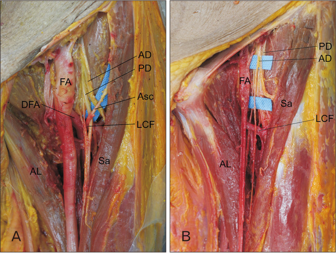
Understanding anatomic variations in neurovascular structure inside the femoral triangle is crucial for regional anesthesiologists performing femoral nerve block. During routine dissection of a cadaver, an ascending branch of the lateral circumflex femoral artery with an anomalous course passing through the femoral nerve, specifically the posterior division, was identified inside the femoral triangle on the left thigh. The novel variation identified in this study occurred in an early stage of prenatal development. Recognition of this anatomic variation will be helpful for reducing unexpected complications during the femoral nerve block and the tensor fascia latae flap. Penetration of the posterior division of the femoral nerve by the arterial branch might cause pain or paresthesia of the medial aspect of the leg in the distribution of the saphenous nerve.
了解股三角内神经血管结构的解剖变异对于实施股神经阻滞的区域麻醉医生至关重要。在一具尸体的常规解剖过程中,在左大腿的股三角内发现了旋股外侧动脉的一个升支,其走行异常,穿过股神经,特别是后股。本研究中发现的这种新变异发生在产前发育的早期阶段。认识到这种解剖变异将有助于减少股神经阻滞和阔筋膜张肌皮瓣手术期间的意外并发症。动脉分支穿透股神经后股可能会导致隐神经分布区域内小腿内侧疼痛或感觉异常。