• 文献检索
  • 文档翻译
  • 深度研究
  • 学术资讯
  • Suppr Zotero 插件Zotero 插件
  • 邀请有礼
  • 套餐&价格
  • 历史记录
应用&插件
Suppr Zotero 插件Zotero 插件浏览器插件Mac 客户端Windows 客户端微信小程序
定价
高级版会员购买积分包购买API积分包
服务
文献检索文档翻译深度研究API 文档MCP 服务
关于我们
关于 Suppr公司介绍联系我们用户协议隐私条款
关注我们

Suppr 超能文献

核心技术专利:CN118964589B侵权必究
粤ICP备2023148730 号-1Suppr @ 2026

文献检索

告别复杂PubMed语法,用中文像聊天一样搜索,搜遍4000万医学文献。AI智能推荐,让科研检索更轻松。

立即免费搜索

文件翻译

保留排版,准确专业,支持PDF/Word/PPT等文件格式,支持 12+语言互译。

免费翻译文档

深度研究

AI帮你快速写综述,25分钟生成高质量综述,智能提取关键信息,辅助科研写作。

立即免费体验

MRI中睾丸结节内纹理过渡特征在鉴别睾丸良恶性病变中的价值

Value of Intra-Perinodular Textural Transition Features from MRI in Distinguishing Between Benign and Malignant Testicular Lesions.

作者信息

Zhang Peipei, Min Xiangde, Feng Zhaoyan, Kang Zhen, Li Basen, Cai Wei, Fan Chanyuan, Yin Xi, Xie Jinke, Lv Wenzhi, Wang Liang

机构信息

Department of Radiology, Tongji Hospital, Tongji Medical College, Huazhong University of Science and Technology, Wuhan 430030, People's Republic of China.

Department of Artificial Intelligence, Julei Technology Company, Wuhan 430030, People's Republic of China.

出版信息

Cancer Manag Res. 2021 Jan 28;13:839-847. doi: 10.2147/CMAR.S288378. eCollection 2021.

DOI:10.2147/CMAR.S288378
PMID:33536790
原文链接:https://pmc.ncbi.nlm.nih.gov/articles/PMC7850382/
Abstract

PURPOSE

To compare the performance of histogram analysis and intra-perinodular textural transition (Ipris) for distinguishing between benign and malignant testicular lesions.

PATIENTS AND METHODS

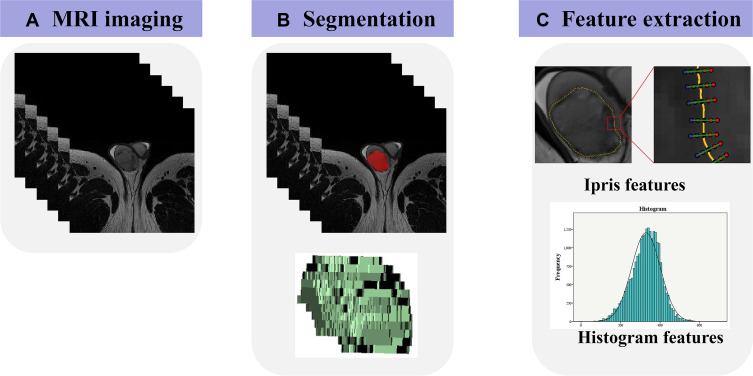
This retrospective study included 76 patients with 80 pathologically confirmed testicular lesions (55 malignant, 25 benign). All patients underwent preoperative T2-weighted imaging (T2WI) on a 3.0T MR scanner. All testicular lesions were manually segmented on axial T2WI, and histogram and Ipris features were extracted. Thirty enrolled patients were randomly selected to estimate the robustness of the features. We used intraclass correlation coefficients (ICCs) to evaluate intra- and interobserver agreement of features, independent -test or Mann-Whitney -test to compare features between benign and malignant lesions, and receiver operating characteristic curve analysis to evaluate the diagnostic performance of features.

RESULTS

Eighteen histogram features and forty-eight Ipris features were extracted from T2WI of each lesion. Most (60/66) histogram and Ipris features had good robustness (ICC of both intra- and interobserver variabilities >0.6). Three histogram and nine Ipris features were significantly different between the benign and malignant groups. The area under the curve values for Energy, TotalEnergy, and Ipris_shell1_id_std were 0.807, 0.808, and 0.708, respectively, which were relatively higher than those of other features.

CONCLUSION

Ipris features may be useful for identifying benign and malignant testicular tumors but have no significant advantage over conventional histogram features.

摘要

目的

比较直方图分析和结节内纹理过渡(Ipris)在鉴别睾丸良恶性病变中的性能。

患者与方法

本回顾性研究纳入了76例患者,共80个经病理证实的睾丸病变(55个恶性,25个良性)。所有患者均在3.0T MR扫描仪上进行了术前T2加权成像(T2WI)。所有睾丸病变均在轴向T2WI上进行手动分割,并提取直方图和Ipris特征。随机选择30例入组患者评估特征的稳健性。我们使用组内相关系数(ICC)来评估特征在观察者内和观察者间的一致性,使用独立样本t检验或曼-惠特尼U检验来比较良恶性病变之间的特征,并使用受试者工作特征曲线分析来评估特征的诊断性能。

结果

从每个病变的T2WI中提取了18个直方图特征和48个Ipris特征。大多数(60/66)直方图和Ipris特征具有良好的稳健性(观察者内和观察者间变异的ICC均>0.6)。良性和恶性组之间有3个直方图特征和9个Ipris特征存在显著差异。能量、总能量和Ipris_shell1_id_std的曲线下面积值分别为0.807、0.808和0.708,相对高于其他特征。

结论

Ipris特征可能有助于鉴别睾丸良恶性肿瘤,但与传统的直方图特征相比没有显著优势。

https://cdn.ncbi.nlm.nih.gov/pmc/blobs/1871/7850382/d2bf10b2d374/CMAR-13-839-g0004.jpg
https://cdn.ncbi.nlm.nih.gov/pmc/blobs/1871/7850382/9c14a095d52b/CMAR-13-839-g0001.jpg
https://cdn.ncbi.nlm.nih.gov/pmc/blobs/1871/7850382/58684dfee910/CMAR-13-839-g0002.jpg
https://cdn.ncbi.nlm.nih.gov/pmc/blobs/1871/7850382/aa230b010454/CMAR-13-839-g0003.jpg
https://cdn.ncbi.nlm.nih.gov/pmc/blobs/1871/7850382/d2bf10b2d374/CMAR-13-839-g0004.jpg

相似文献

1
Value of Intra-Perinodular Textural Transition Features from MRI in Distinguishing Between Benign and Malignant Testicular Lesions.MRI中睾丸结节内纹理过渡特征在鉴别睾丸良恶性病变中的价值
Cancer Manag Res. 2021 Jan 28;13:839-847. doi: 10.2147/CMAR.S288378. eCollection 2021.
2
Discrimination between benign and malignant testicular lesions using volumetric apparent diffusion coefficient histogram analysis.使用容积表观扩散系数直方图分析鉴别睾丸良恶性病变。
Eur J Radiol. 2020 May;126:108939. doi: 10.1016/j.ejrad.2020.108939. Epub 2020 Mar 7.
3
Prediction of Axillary Lymph Node Metastasis in Breast Cancer using Intra-peritumoral Textural Transition Analysis based on Dynamic Contrast-enhanced Magnetic Resonance Imaging.基于动态对比增强磁共振成像的肿瘤内纹理过渡分析预测乳腺癌腋窝淋巴结转移。
Acad Radiol. 2022 Jan;29 Suppl 1:S107-S115. doi: 10.1016/j.acra.2021.02.008. Epub 2021 Mar 10.
4
Characterization of breast masses as benign or malignant at 3.0T MRI with whole-lesion histogram analysis of the apparent diffusion coefficient.利用表观扩散系数的全病变直方图分析在3.0T磁共振成像中对乳腺肿块进行良恶性特征分析。
J Magn Reson Imaging. 2016 Apr;43(4):894-902. doi: 10.1002/jmri.25043. Epub 2015 Sep 7.
5
Can contrast-enhancement computed tomography texture and histogram analyses help to differentiate malignant from benign thyroid nodules?增强 CT 纹理和直方图分析能否帮助鉴别甲状腺良恶性结节?
Jpn J Radiol. 2020 Dec;38(12):1135-1141. doi: 10.1007/s11604-020-01018-z. Epub 2020 Jul 14.
6
Value of conventional magnetic resonance imaging texture analysis in the differential diagnosis of benign and borderline/malignant phyllodes tumors of the breast.常规磁共振成像纹理分析在鉴别诊断乳腺良性和交界性/恶性叶状肿瘤中的价值。
Cancer Imaging. 2021 Mar 12;21(1):29. doi: 10.1186/s40644-021-00398-3.
7
Application of Texture Analysis Based on Sagittal Fat-Suppression and Oblique Axial T2-Weighted Magnetic Resonance Imaging to Identify Lymph Node Invasion Status of Rectal Cancer.基于矢状位脂肪抑制和斜轴位T2加权磁共振成像的纹理分析在直肠癌淋巴结侵犯状态识别中的应用
Front Oncol. 2020 Aug 7;10:1364. doi: 10.3389/fonc.2020.01364. eCollection 2020.
8
Discriminating malignant from benign testicular masses using machine-learning based radiomics signature of appearance diffusion coefficient maps: Comparing with conventional mean and minimum ADC values.基于机器学习的表观扩散系数图纹理特征鉴别睾丸良恶性肿块:与常规平均和最小 ADC 值的比较。
Eur J Radiol. 2022 Mar;148:110158. doi: 10.1016/j.ejrad.2022.110158. Epub 2022 Jan 15.
9
Diagnostic performance of multi-parametric MRI to differentiate benign sex cord stromal tumors from malignant (non-stromal and stromal) testicular neoplasms.多参数 MRI 鉴别良恶性(非间质和间质)睾丸性索间质肿瘤的诊断性能。
Abdom Radiol (NY). 2021 Jan;46(1):319-330. doi: 10.1007/s00261-020-02621-4. Epub 2020 Jun 22.
10
Solid bone tumors of the spine: Diagnostic performance of apparent diffusion coefficient measured using diffusion-weighted MRI using histology as a reference standard.脊柱骨肿瘤:以组织学为参考标准的磁共振扩散加权成像测量表观扩散系数的诊断性能。
J Magn Reson Imaging. 2018 Apr;47(4):1034-1042. doi: 10.1002/jmri.25826. Epub 2017 Jul 29.

引用本文的文献

1
An Overview of the Role of Multiparametric MRI in the Investigation of Testicular Tumors.多参数磁共振成像在睾丸肿瘤研究中的作用概述
Cancers (Basel). 2022 Aug 13;14(16):3912. doi: 10.3390/cancers14163912.
2
Contemporary options and future perspectives: three examples highlighting the challenges in testicular cancer imaging.当代选择与未来展望:三个突出睾丸癌成像挑战的实例。
World J Urol. 2022 Feb;40(2):307-315. doi: 10.1007/s00345-021-03856-6. Epub 2021 Nov 15.
3
When to ask for an MRI of the scrotum.何时需要进行阴囊 MRI 检查。

本文引用的文献

1
Diagnostic performance of multi-parametric MRI to differentiate benign sex cord stromal tumors from malignant (non-stromal and stromal) testicular neoplasms.多参数 MRI 鉴别良恶性(非间质和间质)睾丸性索间质肿瘤的诊断性能。
Abdom Radiol (NY). 2021 Jan;46(1):319-330. doi: 10.1007/s00261-020-02621-4. Epub 2020 Jun 22.
2
Discrimination between benign and malignant testicular lesions using volumetric apparent diffusion coefficient histogram analysis.使用容积表观扩散系数直方图分析鉴别睾丸良恶性病变。
Eur J Radiol. 2020 May;126:108939. doi: 10.1016/j.ejrad.2020.108939. Epub 2020 Mar 7.
3
Texture Analysis of Multi-Shot Echo-planar Diffusion-Weighted Imaging in Head and Neck Squamous Cell Carcinoma: The Diagnostic Value for Nodal Metastasis.
Andrology. 2021 Sep;9(5):1395-1409. doi: 10.1111/andr.13032. Epub 2021 Jun 11.
头颈部鳞状细胞癌多次激发回波平面扩散加权成像的纹理分析:对淋巴结转移的诊断价值
J Clin Med. 2019 Oct 23;8(11):1767. doi: 10.3390/jcm8111767.
4
Stability and reproducibility of computed tomography radiomic features extracted from peritumoral regions of lung cancer lesions.从肺癌病变瘤周区域提取的 CT 放射组学特征的稳定性和可重复性。
Med Phys. 2019 Nov;46(11):5075-5085. doi: 10.1002/mp.13808. Epub 2019 Sep 23.
5
Rectal cancer: can T2WI histogram of the primary tumor help predict the existence of lymph node metastasis?直肠癌:原发肿瘤 T2WI 直方图能否帮助预测淋巴结转移的存在?
Eur Radiol. 2019 Dec;29(12):6469-6476. doi: 10.1007/s00330-019-06328-z. Epub 2019 Jul 5.
6
Testicular germ cell tumor: a comprehensive review.睾丸生殖细胞肿瘤:全面综述。
Cell Mol Life Sci. 2019 May;76(9):1713-1727. doi: 10.1007/s00018-019-03022-7. Epub 2019 Jan 22.
7
The role of tumor size, ultrasonographic findings, and serum tumor markers in predicting the likelihood of malignant testicular histology.肿瘤大小、超声表现和血清肿瘤标志物在预测睾丸组织恶性程度中的作用。
Asian J Androl. 2019 Mar-Apr;21(2):196-200. doi: 10.4103/aja.aja_119_18.
8
MRI of testicular malignancies.MRI 检查在睾丸恶性肿瘤中的应用。
Abdom Radiol (NY). 2019 Mar;44(3):1070-1082. doi: 10.1007/s00261-018-1816-5.
9
Testicular cancer.睾丸癌。
Nat Rev Dis Primers. 2018 Oct 5;4(1):29. doi: 10.1038/s41572-018-0029-0.
10
Computed tomography-based texture analysis of bladder cancer: differentiating urothelial carcinoma from micropapillary carcinoma.基于计算机断层扫描的膀胱癌纹理分析:鉴别尿路上皮癌与微乳头状癌。
Abdom Radiol (NY). 2019 Jan;44(1):201-208. doi: 10.1007/s00261-018-1694-x.