van Genderen Anne Metje, Jansen Katja, Kristen Marleen, van Duijn Joost, Li Yang, Schuurmans Carl C L, Malda Jos, Vermonden Tina, Jansen Jitske, Masereeuw Rosalinde, Castilho Miguel
Division of Pharmacology, Utrecht Institute for Pharmaceutical Sciences, Utrecht University, Utrecht, Netherlands.
Department of Orthopaedics, University Medical Center Utrecht, Utrecht University, Utrecht, Netherlands.
Front Bioeng Biotechnol. 2021 Jan 18;8:617364. doi: 10.3389/fbioe.2020.617364. eCollection 2020.
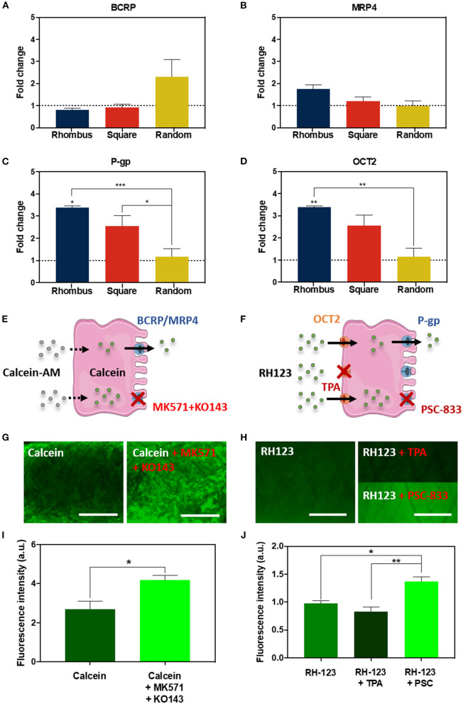
To date, tubular tissue engineering relies on large, non-porous tubular scaffolds (Ø > 2 mm) for mechanical self-support, or smaller (Ø 150-500 μm) tubes within bulk hydrogels for studying renal transport phenomena. To advance the engineering of kidney tubules for future implantation, constructs should be both self-supportive and yet small-sized and highly porous. Here, we hypothesize that the fabrication of small-sized porous tubular scaffolds with a highly organized fibrous microstructure by means of melt-electrowriting (MEW) allows the development of self-supported kidney proximal tubules with enhanced properties. A custom-built melt-electrowriting (MEW) device was used to fabricate tubular fibrous scaffolds with small diameter sizes (Ø = 0.5, 1, 3 mm) and well-defined, porous microarchitectures (rhombus, square, and random). Human umbilical vein endothelial cells (HUVEC) and human conditionally immortalized proximal tubular epithelial cells (ciPTEC) were seeded into the tubular scaffolds and tested for monolayer formation, integrity, and organization, as well as for extracellular matrix (ECM) production and renal transport functionality. Tubular fibrous scaffolds were successfully manufactured by fine control of MEW instrument parameters. A minimum inner diameter of 1 mm and pore sizes of 0.2 mm were achieved and used for subsequent cell experiments. While HUVEC were unable to bridge the pores, ciPTEC formed tight monolayers in all scaffold microarchitectures tested. Well-defined rhombus-shaped pores outperformed and facilitated unidirectional cell orientation, increased collagen type IV deposition, and expression of the renal transporters and differentiation markers organic cation transporter 2 (OCT2) and P-glycoprotein (P-gp). Here, we present smaller diameter engineered kidney tubules with microgeometry-directed cell functionality. Due to the well-organized tubular fiber scaffold microstructure, the tubes are mechanically self-supported, and the self-produced ECM constitutes the only barrier between the inner and outer compartment, facilitating rapid and active solute transport.
迄今为止,管状组织工程依赖于大型、无孔的管状支架(直径大于2毫米)以实现机械自支撑,或者依赖于块状水凝胶中较小(直径150 - 500微米)的管子来研究肾脏转运现象。为了推进未来可植入的肾小管工程,构建物应既具有自支撑性,又要尺寸小且高度多孔。在此,我们假设通过熔体静电纺丝(MEW)制造具有高度有序纤维微观结构的小型多孔管状支架,能够开发出具有增强性能的自支撑肾近端小管。使用定制的熔体静电纺丝(MEW)装置制造具有小直径尺寸(直径 = 0.5、1、3毫米)和明确多孔微结构(菱形、方形和随机)的管状纤维支架。将人脐静脉内皮细胞(HUVEC)和人条件永生化近端肾小管上皮细胞(ciPTEC)接种到管状支架中,并测试其单层形成、完整性和组织性,以及细胞外基质(ECM)产生和肾脏转运功能。通过对MEW仪器参数的精细控制,成功制造出管状纤维支架。实现了最小内径1毫米和孔径0.2毫米,并用于后续细胞实验。虽然HUVEC无法跨越孔隙,但ciPTEC在所有测试的支架微结构中都形成了紧密的单层。明确的菱形孔隙表现更优,促进了细胞的单向定向排列,增加了IV型胶原蛋白的沉积,以及肾转运蛋白和分化标志物有机阳离子转运体2(OCT2)和P - 糖蛋白(P - gp)的表达。在此,我们展示了具有微几何结构导向细胞功能的小直径工程化肾小管。由于管状纤维支架微观结构组织良好,这些管子在机械上是自支撑的,并且自我产生的ECM构成了内外腔室之间的唯一屏障,促进了快速且活跃的溶质转运。