Department of Radiology, the First Affiliated Hospital of Xi'an Jiaotong University, 277 West Yanta Rd, Xi'an, 710061, Shaanxi, China.
Department of Radiology, University of Wisconsin-Madison, Room 7137, 1111 Highland Ave, Madison, WI, 53705, USA.
Eur J Nucl Med Mol Imaging. 2021 Aug;48(9):2737-2748. doi: 10.1007/s00259-021-05216-3. Epub 2021 Feb 3.
We dual-labeled an intercellular adhesion molecule-1 (ICAM-1) monoclonal antibody (mAb) and evaluated its effectiveness for lesion detection and surgical navigation in pancreatic ductal adenocarcinoma (PDAC) via multiple noninvasive imaging approaches, including positron emission tomography (PET), near-infrared fluorescence (NIRF), and Cerenkov luminescence imaging (CLI).
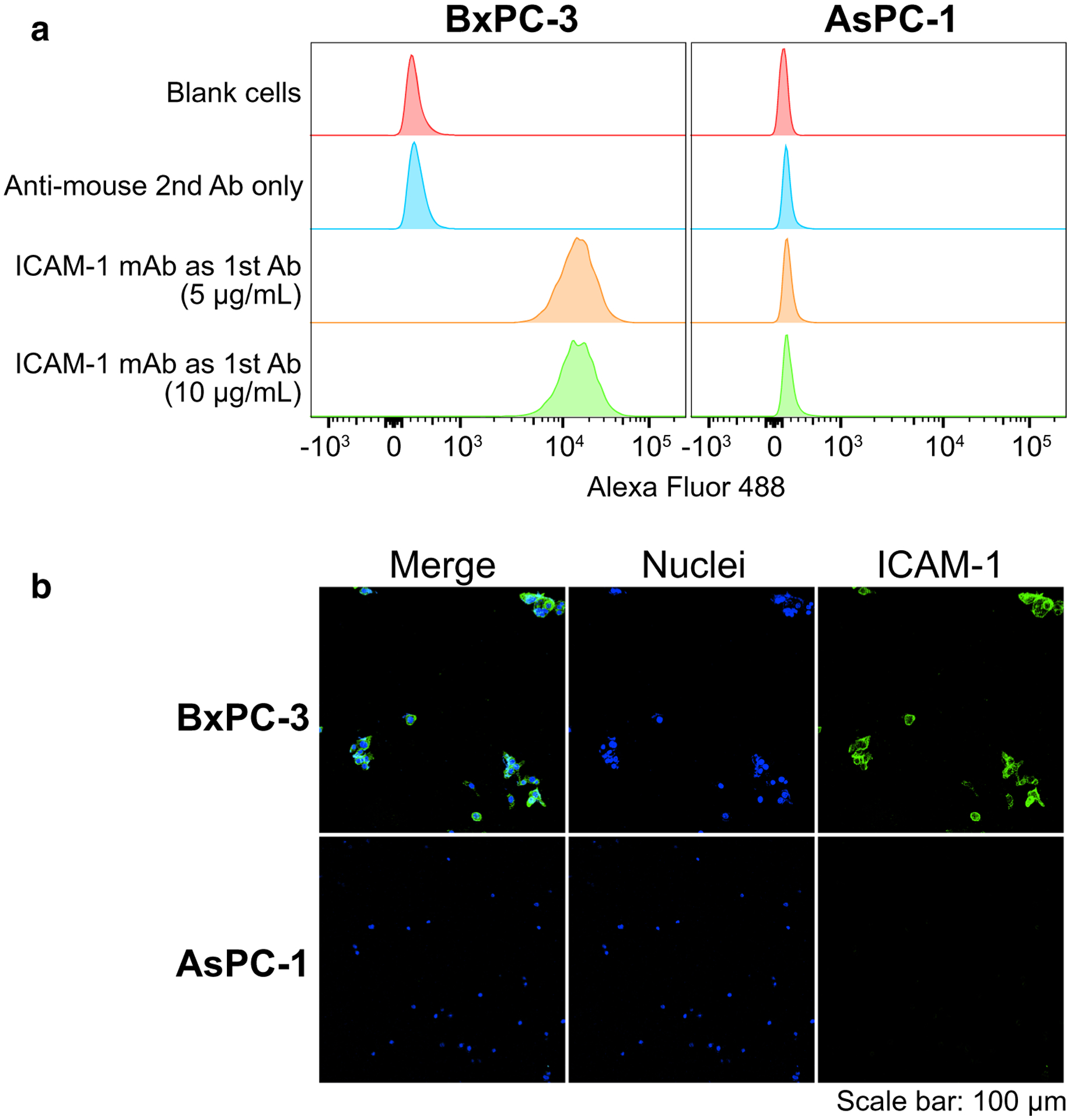
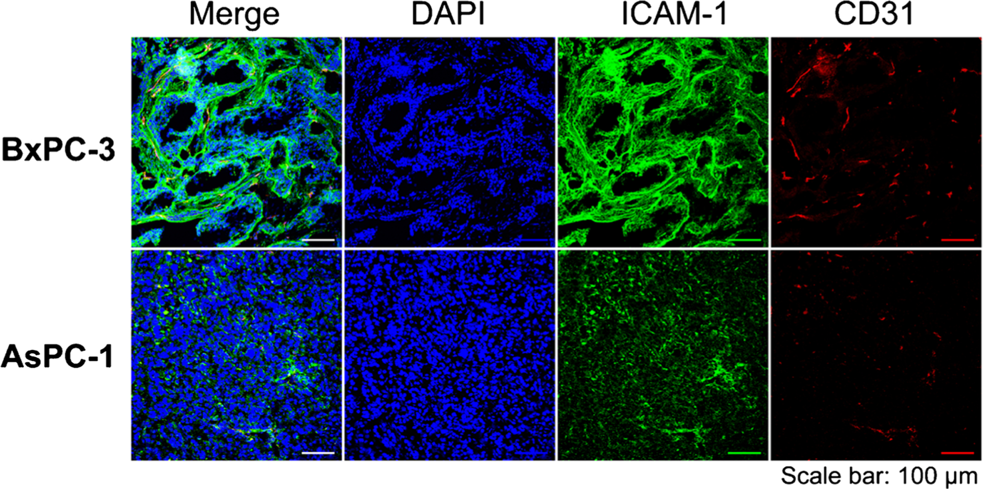
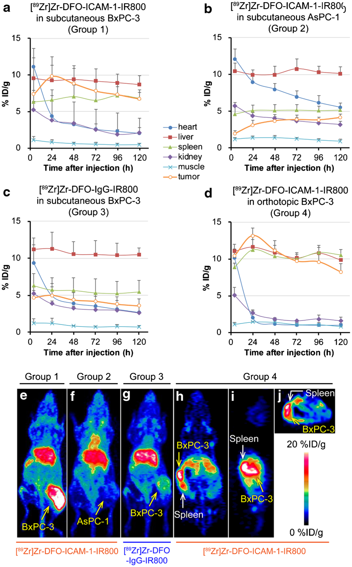
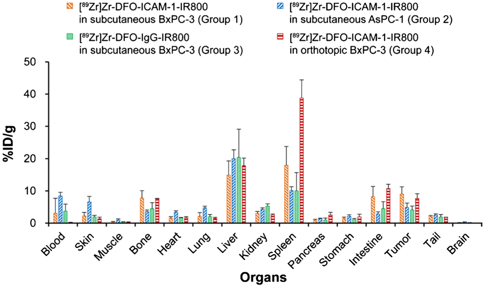
ICAM-1 expression in PDAC cell lines (BxPC-3 and AsPC-1) was assessed via flow cytometry and immunofluorescent staining. An ICAM-1 mAb labeled by IRDye 800CW and radionuclide zirconium-89 (denoted as [Zr]Zr-DFO-ICAM-1-IR800) was synthesized. Its performance was validated via in vivo comparative PET/NIRF/CLI and biodistribution (Bio-D) studies in nude mice bearing subcutaneous BxPC-3/AsPC-1 tumors or orthotopic BxPC-3 tumor models using nonspecific IgG as an isotype control tracer.
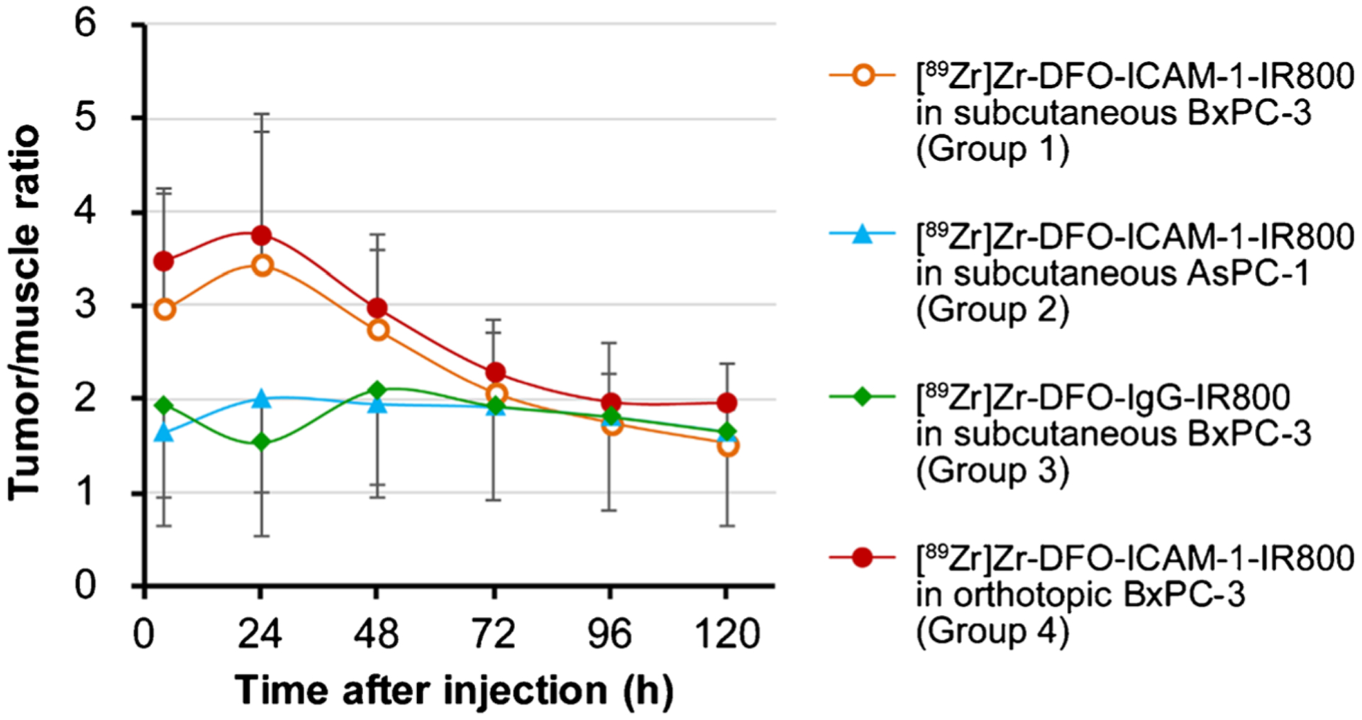
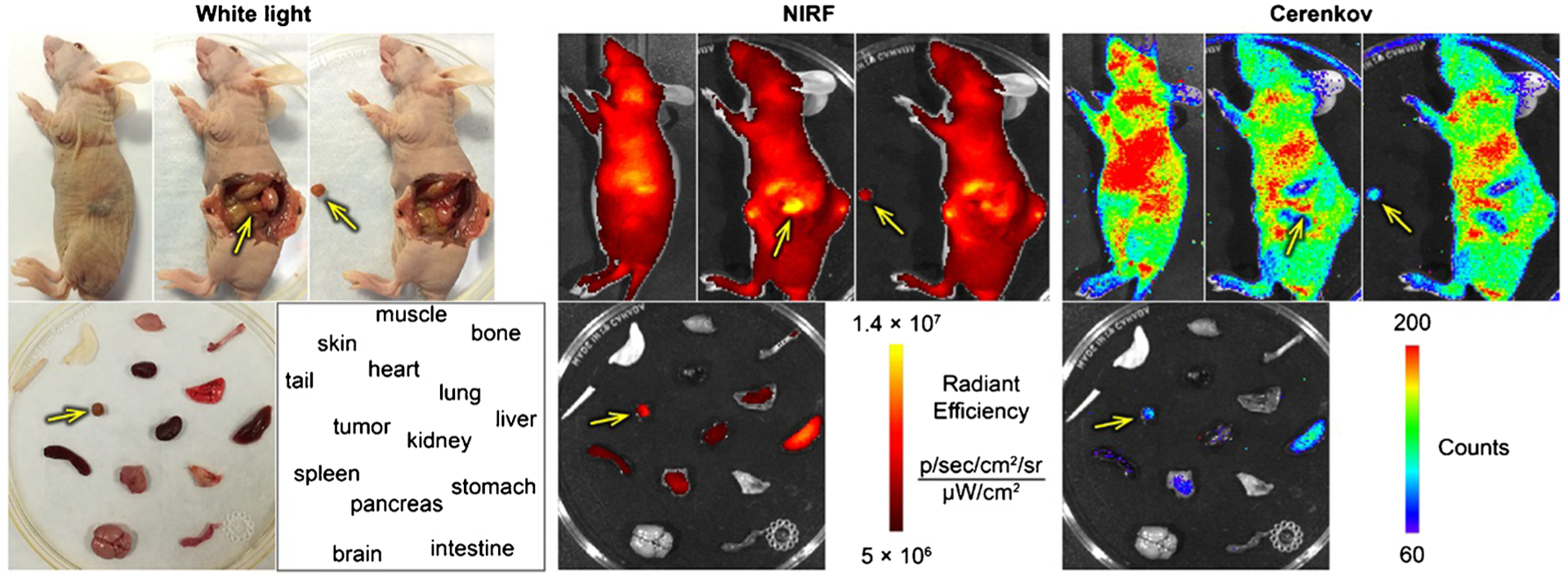
ICAM-1 expression was strong in the BxPC-3 and minimal in the AsPC-1 cell line. Both multimodality imaging and Bio-D data exhibited more prominent uptake of [Zr]Zr-DFO-ICAM-1-IR800 in BxPC-3 tumors than in AsPC-1 tumors. The uptake of [Zr]Zr-DFO-IgG-IR800 in BxPC-3 tumors was similar to that of [Zr]Zr-DFO-ICAM-1-IR800 in AsPC-1 tumors. These results demonstrate the desirable affinity and specificity of [Zr]Zr-DFO-ICAM-1-IR800 compared to [Zr]Zr-DFO-IgG-IR800. Orthotopic BxPC-3 tumor foci could also be clearly delineated by [Zr]Zr-DFO-ICAM-1-IR800. An intermodal match was achieved in the ICAM-1-targeted immunoPET/NIRF/CLI. The positive expression levels of ICAM-1 in BxPC-3 tumor tissue were further confirmed by immunohistopathology.
We successfully developed a dual-labeled ICAM-1-targeted tracer for PET/NIRF/CLI of PDAC that can facilitate better diagnosis and intervention of PDAC upon clinical translation.
我们对细胞间黏附分子-1(ICAM-1)单克隆抗体(mAb)进行双标记,并通过多种非侵入性成像方法,包括正电子发射断层扫描(PET)、近红外荧光(NIRF)和切伦科夫发光成像(CLI),评估其在胰腺导管腺癌(PDAC)中的病灶检测和手术导航的有效性。
通过流式细胞术和免疫荧光染色评估 PDAC 细胞系(BxPC-3 和 AsPC-1)中 ICAM-1 的表达。合成了一种通过 IRDye 800CW 和放射性核素锆-89 标记的 ICAM-1 mAb(表示为[Zr]Zr-DFO-ICAM-1-IR800)。通过在皮下接种 BxPC-3/AsPC-1 肿瘤或原位 BxPC-3 肿瘤模型的裸鼠中进行体内比较 PET/NIRF/Cli 和生物分布(Bio-D)研究,以非特异性 IgG 作为同种型对照示踪剂,验证其性能。
ICAM-1 在 BxPC-3 细胞系中的表达较强,在 AsPC-1 细胞系中的表达较弱。多模态成像和 Bio-D 数据均显示,[Zr]Zr-DFO-ICAM-1-IR800 在 BxPC-3 肿瘤中的摄取明显高于 AsPC-1 肿瘤。BxPC-3 肿瘤中[Zr]Zr-DFO-IgG-IR800 的摄取与 AsPC-1 肿瘤中[Zr]Zr-DFO-ICAM-1-IR800 的摄取相似。这些结果表明,与[Zr]Zr-DFO-IgG-IR800 相比,[Zr]Zr-DFO-ICAM-1-IR800 具有理想的亲和力和特异性。[Zr]Zr-DFO-ICAM-1-IR800 还可以清晰地描绘出原位 BxPC-3 肿瘤灶。免疫 PET/NIRF/Cli 实现了模态间匹配。免疫组织病理学进一步证实了 BxPC-3 肿瘤组织中 ICAM-1 的阳性表达水平。
我们成功开发了一种用于 PDAC 的双标记 ICAM-1 靶向示踪剂,可在临床转化后促进 PDAC 的更好诊断和干预。