• 文献检索
  • 文档翻译
  • 深度研究
  • 学术资讯
  • Suppr Zotero 插件Zotero 插件
  • 邀请有礼
  • 套餐&价格
  • 历史记录
应用&插件
Suppr Zotero 插件Zotero 插件浏览器插件Mac 客户端Windows 客户端微信小程序
定价
高级版会员购买积分包购买API积分包
服务
文献检索文档翻译深度研究API 文档MCP 服务
关于我们
关于 Suppr公司介绍联系我们用户协议隐私条款
关注我们

Suppr 超能文献

核心技术专利:CN118964589B侵权必究
粤ICP备2023148730 号-1Suppr @ 2026

文献检索

告别复杂PubMed语法,用中文像聊天一样搜索,搜遍4000万医学文献。AI智能推荐,让科研检索更轻松。

立即免费搜索

文件翻译

保留排版,准确专业,支持PDF/Word/PPT等文件格式,支持 12+语言互译。

免费翻译文档

深度研究

AI帮你快速写综述,25分钟生成高质量综述,智能提取关键信息,辅助科研写作。

立即免费体验

超微肺囊肿在淋巴管平滑肌瘤病中不阻塞气流而损害弥散。

Ultra-Small Lung Cysts Impair Diffusion Without Obstructing Air Flow in Lymphangioleiomyomatosis.

机构信息

National Heart, Lung, and Blood Institute, National Institutes of Health, Bethesda, MD.

National Heart, Lung, and Blood Institute, National Institutes of Health, Bethesda, MD.

出版信息

Chest. 2021 Jul;160(1):199-208. doi: 10.1016/j.chest.2021.01.077. Epub 2021 Feb 5.

DOI:10.1016/j.chest.2021.01.077
PMID:33549601
原文链接:https://pmc.ncbi.nlm.nih.gov/articles/PMC8295912/
Abstract

BACKGROUND

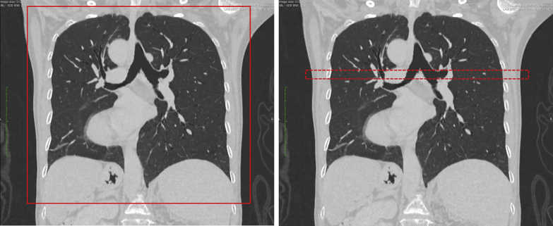
Lymphangioleiomyomatosis (LAM) is a rare lung disease found primarily in women of childbearing age, characterized by the formation of air-filled cysts, which may be associated with reductions in lung function. An experimental, regional ultra-high resolution CT scan identified an additional volume of cysts relative to standard chest CT imaging, which consisted primarily of ultra-small cysts.

RESEARCH QUESTION

What is the impact of these ultra-small cysts on the pulmonary function of patients with LAM?

STUDY DESIGN AND METHODS

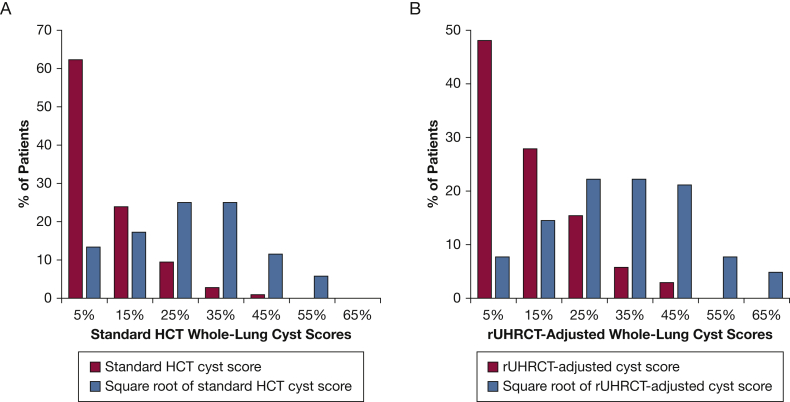
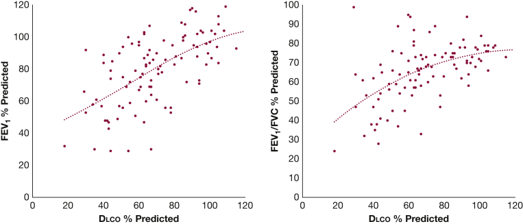
A group of 103 patients with LAM received pulmonary function tests and a CT examination in the same visit. Cyst score, the percentage lung volume occupied by cysts, was measured by using commercial software approved by the US Food and Drug Administration. The association between cyst scores and pulmonary function tests of diffusing capacity of the lungs for carbon monoxide (Dlco) (% predicted), FEV (% predicted), and FEV/FVC (% predicted) was assessed with statistical analysis adjusted for demographic variables. The distributions of average cyst size and ultra-small cyst fraction among the patients were evaluated.

RESULTS

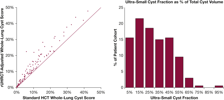
The additional cyst volume identified by the experimental, higher resolution scan consisted of cysts of 2.2 ± 0.8 mm diameter on average and are thus labeled the "ultra-small cyst fraction." It accounted for 27.9 ± 19.0% of the total cyst volume among the patients. The resulting adjusted, whole-lung cyst scores better explained the variance of Dlco (P < .001 adjusted for multiple comparisons) but not FEV and FEV/FVC (P = 1.00). The ultra-small cyst fraction contributed to the reduction in Dlco (P < .001) but not to FEV and FEV/FVC (P = .760 and .575, respectively). The ultra-small cyst fraction and average cyst size were correlated with cyst burden, FEV, and FEV/FVC but less with Dlco.

INTERPRETATION

The ultra-small cysts primarily contributed to the reduction in Dlco, with minimal effects on FEV and FEV/FVC. Patients with lower cyst burden and better FEV and FEV/FVC tended to have smaller average cyst size and higher ultra-small cyst fraction.

CLINICAL TRIAL REGISTRATION

ClinicalTrials.gov; No.: NCT00001465; URL: www.clinicaltrials.gov.

摘要

背景

淋巴管平滑肌瘤病(LAM)是一种主要发生在育龄妇女中的罕见肺部疾病,其特征是形成充满空气的囊肿,这可能与肺功能下降有关。一项实验性的区域性超高分辨率 CT 扫描发现,与标准胸部 CT 成像相比,囊肿的体积增加,主要由超小囊肿组成。

研究问题

这些超小囊肿对 LAM 患者的肺功能有什么影响?

研究设计和方法

一组 103 名 LAM 患者在同一次就诊时接受了肺功能检查和 CT 检查。使用美国食品和药物管理局批准的商业软件测量囊肿评分,即囊肿占肺体积的百分比。通过统计分析评估囊肿评分与弥散量一氧化碳(Dlco)(%预计值)、FEV(%预计值)和 FEV/FVC(%预计值)之间的相关性,该分析调整了人口统计学变量。评估了患者之间平均囊肿大小和超小囊肿分数的分布。

结果

实验性更高分辨率扫描所识别的额外囊肿体积由平均直径为 2.2±0.8mm 的囊肿组成,因此被标记为“超小囊肿分数”。它占患者总囊肿体积的 27.9±19.0%。调整后的全肺囊肿评分更好地解释了 Dlco 的变化(P<0.001,经多次比较调整),但不能解释 FEV 和 FEV/FVC 的变化(P=1.00)。超小囊肿分数与 Dlco 降低有关(P<0.001),但与 FEV 和 FEV/FVC 无关(P=0.760 和 0.575)。超小囊肿分数和平均囊肿大小与囊肿负担、FEV 和 FEV/FVC 相关,但与 Dlco 相关性较低。

结论

超小囊肿主要导致 Dlco 降低,对 FEV 和 FEV/FVC 的影响较小。囊肿负担较低、FEV 和 FEV/FVC 较好的患者,其平均囊肿大小较小,超小囊肿分数较高。

临床试验注册

ClinicalTrials.gov;编号:NCT00001465;网址:www.clinicaltrials.gov。

https://cdn.ncbi.nlm.nih.gov/pmc/blobs/56e2/8295912/490cf00c728b/gr7.jpg
https://cdn.ncbi.nlm.nih.gov/pmc/blobs/56e2/8295912/9311a67b6f93/gr1.jpg
https://cdn.ncbi.nlm.nih.gov/pmc/blobs/56e2/8295912/4714aa057d15/gr2.jpg
https://cdn.ncbi.nlm.nih.gov/pmc/blobs/56e2/8295912/6dc6b33b578d/gr3.jpg
https://cdn.ncbi.nlm.nih.gov/pmc/blobs/56e2/8295912/09a015b2231b/gr4.jpg
https://cdn.ncbi.nlm.nih.gov/pmc/blobs/56e2/8295912/ca0e79515bc2/gr5.jpg
https://cdn.ncbi.nlm.nih.gov/pmc/blobs/56e2/8295912/02d64585b73d/gr6.jpg
https://cdn.ncbi.nlm.nih.gov/pmc/blobs/56e2/8295912/490cf00c728b/gr7.jpg

相似文献

1
Ultra-Small Lung Cysts Impair Diffusion Without Obstructing Air Flow in Lymphangioleiomyomatosis.超微肺囊肿在淋巴管平滑肌瘤病中不阻塞气流而损害弥散。
Chest. 2021 Jul;160(1):199-208. doi: 10.1016/j.chest.2021.01.077. Epub 2021 Feb 5.
2
Cyst Ventilation Heterogeneity and Alveolar Airspace Dilation as Early Disease Markers in Lymphangioleiomyomatosis.淋巴管平滑肌瘤病的囊泡通气异质性和肺泡气腔扩张作为早期疾病标志物。
Ann Am Thorac Soc. 2019 Aug;16(8):1008-1016. doi: 10.1513/AnnalsATS.201812-880OC.
3
Computer grading of lung disease severity in patients with lymphangioleiomyomatosis referred for transplantation.淋巴管肌瘤病患者移植前肺疾病严重程度的计算机评分。
BMC Pulm Med. 2022 Sep 24;22(1):362. doi: 10.1186/s12890-022-02123-7.
4
Effects of sirolimus in lymphangioleiomyomatosis patients on lung cysts and pulmonary function: long-term follow-up observational study.淋巴管肌瘤病患者使用西罗莫司对肺囊肿和肺功能的影响:长期随访观察性研究。
Eur Radiol. 2020 Feb;30(2):735-743. doi: 10.1007/s00330-019-06412-4. Epub 2019 Aug 30.
5
Pulmonary lymphangioleiomyomatosis. A study of 69 patients. Groupe d'Etudes et de Recherche sur les Maladies "Orphelines" Pulmonaires (GERM"O"P).肺淋巴管平滑肌瘤病。69例患者的研究。肺部“孤儿”疾病研究与探索小组(GERM"O"P)。
Medicine (Baltimore). 1999 Sep;78(5):321-37. doi: 10.1097/00005792-199909000-00004.
6
Cyst-based measurements for assessing lymphangioleiomyomatosis in computed tomography.基于囊肿的计算机断层扫描测量用于评估淋巴管平滑肌瘤病
Med Phys. 2015 May;42(5):2287-95. doi: 10.1118/1.4916655.
7
Clinical CT underestimation of the percentage volume occupied by cysts in patients with lymphangioleiomyomatosis.淋巴管肌瘤病患者 CT 临床低估囊肿体积百分比。
Clin Imaging. 2020 Feb;59(2):119-125. doi: 10.1016/j.clinimag.2019.11.004. Epub 2019 Nov 27.
8
Chest CT Scan at Radiation Dose of a Posteroanterior and Lateral Chest Radiograph Series: A Proof of Principle in Lymphangioleiomyomatosis.胸部 CT 扫描在前后位和侧位胸部 X 线摄影系列中的辐射剂量:淋巴管平滑肌瘤病的原理证明。
Chest. 2019 Mar;155(3):528-533. doi: 10.1016/j.chest.2018.09.007. Epub 2018 Oct 3.
9
Automated algorithm for quantifying the extent of cystic change on volumetric chest CT: initial results in Lymphangioleiomyomatosis.用于量化胸部容积CT上囊性改变范围的自动化算法:淋巴管平滑肌瘤病的初步结果
AJR Am J Roentgenol. 2009 Apr;192(4):1037-44. doi: 10.2214/AJR.07.3334.
10
Use of CT Imaging to Quantify Progression and Response to Treatment in Lymphangioleiomyomatosis.使用 CT 成像定量评估淋巴管平滑肌瘤病的进展和治疗反应。
Chest. 2019 May;155(5):962-971. doi: 10.1016/j.chest.2019.01.004. Epub 2019 Jan 17.

引用本文的文献

1
Cystic Lung Phantom to Validate Clinical CT Protocols.用于验证临床CT方案的囊性肺模体
Methods Protoc. 2025 Jun 13;8(3):63. doi: 10.3390/mps8030063.
2
Cystic Lung Phantom to Validate Clinical CT Protocols.用于验证临床CT方案的囊性肺模体
ArXiv. 2025 Mar 25:arXiv:2503.19747v1.
3
Clinical Trial Validation of Automated Segmentation and Scoring of Pulmonary Cysts in Thoracic CT Scans.胸部CT扫描中肺囊肿自动分割与评分的临床试验验证

本文引用的文献

1
Quantitative Analysis of Cystic Lung Diseases by Use of Paired Inspiratory and Expiratory CT: Estimation of the Extent of Cyst-Airway Communication and Evaluation of Diagnostic Utility.使用吸气期与呼气期配对CT对肺囊性疾病进行定量分析:评估囊肿与气道的连通程度及诊断效用
Radiol Cardiothorac Imaging. 2020 Apr 30;2(2):e190097. doi: 10.1148/ryct.2020190097. eCollection 2020 Apr.
2
Diffusing Capacity Is an Independent Predictor of Outcomes in Pulmonary Hypertension Associated With COPD.弥散能力是慢性阻塞性肺疾病相关肺动脉高压预后的独立预测指标。
Chest. 2020 Aug;158(2):722-734. doi: 10.1016/j.chest.2020.02.047. Epub 2020 Mar 14.
3
Diagnostics (Basel). 2024 Jul 15;14(14):1529. doi: 10.3390/diagnostics14141529.
4
Images in Vascular Medicine: High-resolution CT imaging of arterial calcification in the hands and legs of patients with CD73 deficiency.血管医学影像:CD73缺乏患者手部和腿部动脉钙化的高分辨率CT成像
Vasc Med. 2024 Jun;29(3):342-344. doi: 10.1177/1358863X231214711. Epub 2023 Dec 22.
5
Automated Segmentation and Measurements of Pulmonary Cysts in Lymphangioleiomyomatosis across Multiple CT Scanner Platforms over a Period of Two Decades.二十年间跨多个CT扫描平台对淋巴管平滑肌瘤病中肺囊肿的自动分割与测量
Bioengineering (Basel). 2023 Oct 27;10(11):1255. doi: 10.3390/bioengineering10111255.
6
Lymphangioleiomyomatosis: circulating levels of FGF23 and pulmonary diffusion.淋巴管平滑肌瘤病:循环中 FGF23 水平与肺弥散功能。
J Bras Pneumol. 2023 May 1;49(2):e20220356. doi: 10.36416/1806-3756/e20220356. eCollection 2023.
7
Novel Pulmonary Cyst Characteristics Associated with the Diffusing Capacity of the Lung in Lymphangioleiomyomatosis: A Cross-Sectional Clinical Trial.与淋巴管平滑肌瘤病肺弥散功能相关的新型肺囊肿特征:一项横断面临床试验
Ann Am Thorac Soc. 2023 Jul;20(7):1073-1076. doi: 10.1513/AnnalsATS.202212-1003RL.
8
Computer grading of lung disease severity in patients with lymphangioleiomyomatosis referred for transplantation.淋巴管肌瘤病患者移植前肺疾病严重程度的计算机评分。
BMC Pulm Med. 2022 Sep 24;22(1):362. doi: 10.1186/s12890-022-02123-7.
Clinical CT underestimation of the percentage volume occupied by cysts in patients with lymphangioleiomyomatosis.
淋巴管肌瘤病患者 CT 临床低估囊肿体积百分比。
Clin Imaging. 2020 Feb;59(2):119-125. doi: 10.1016/j.clinimag.2019.11.004. Epub 2019 Nov 27.
4
Diffusing Capacity of Carbon Monoxide in Assessment of COPD.一氧化碳弥散量在 COPD 评估中的应用。
Chest. 2019 Dec;156(6):1111-1119. doi: 10.1016/j.chest.2019.06.035. Epub 2019 Jul 25.
5
Cyst Ventilation Heterogeneity and Alveolar Airspace Dilation as Early Disease Markers in Lymphangioleiomyomatosis.淋巴管平滑肌瘤病的囊泡通气异质性和肺泡气腔扩张作为早期疾病标志物。
Ann Am Thorac Soc. 2019 Aug;16(8):1008-1016. doi: 10.1513/AnnalsATS.201812-880OC.
6
Use of CT Imaging to Quantify Progression and Response to Treatment in Lymphangioleiomyomatosis.使用 CT 成像定量评估淋巴管平滑肌瘤病的进展和治疗反应。
Chest. 2019 May;155(5):962-971. doi: 10.1016/j.chest.2019.01.004. Epub 2019 Jan 17.
7
Emphysema quantification using chest CT: influence of radiation dose reduction and reconstruction technique.使用胸部CT进行肺气肿定量分析:辐射剂量降低和重建技术的影响
Eur Radiol Exp. 2018 Nov 7;2(1):30. doi: 10.1186/s41747-018-0064-3.
8
Chest CT Scan at Radiation Dose of a Posteroanterior and Lateral Chest Radiograph Series: A Proof of Principle in Lymphangioleiomyomatosis.胸部 CT 扫描在前后位和侧位胸部 X 线摄影系列中的辐射剂量:淋巴管平滑肌瘤病的原理证明。
Chest. 2019 Mar;155(3):528-533. doi: 10.1016/j.chest.2018.09.007. Epub 2018 Oct 3.
9
Optimization of a secondary VOI protocol for lung imaging in a clinical CT scanner.临床CT扫描仪中肺部成像的二次感兴趣区(VOI)协议优化
J Appl Clin Med Phys. 2018 Jul;19(4):271-280. doi: 10.1002/acm2.12354. Epub 2018 May 21.
10
Difference of the progression of pulmonary cysts assessed by computed tomography among COPD, lymphangioleiomyomatosis, and Birt-Hogg-Dubé syndrome.慢性阻塞性肺疾病、淋巴管平滑肌瘤病和 Birt-Hogg-Dubé 综合征患者 CT 评估肺囊肿进展的差异。
PLoS One. 2017 Dec 8;12(12):e0188771. doi: 10.1371/journal.pone.0188771. eCollection 2017.