Division of Gastroenterology, Hepatology and EndoscopyBrigham and Women's HospitalBostonMAUSA.
Harvard Medical SchoolBostonMAUSA.
Hepatol Commun. 2020 Nov 21;5(2):283-292. doi: 10.1002/hep4.1635. eCollection 2021 Feb.
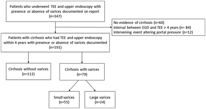
Despite scant evidence, current guidelines indicate that esophageal varices are a relative contraindication to transesophageal echocardiography (TEE). The aim of this study is to compare the risk of gastrointestinal bleeding following TEE among cirrhotic patients with and without endoscopically-documented esophageal varices. This is a retrospective analysis of patients with cirrhosis who underwent upper endoscopy within 4 years of TEE at five institutions between January 2000 and March 2020. Primary outcome was overt gastrointestinal bleeding. Secondary outcomes were hemoglobin decline by at least 2 g/dL or blood transfusion within 48 hours following TEE. Of the 191 patients, 79 (41.4%) had esophageal varices (30.4% large). No patient experienced a primary outcome. Secondary outcomes occurred in 52 (27.2%): 28 (35.4%) with esophageal varices and 24 (21.4%) without varices. After propensity-score covariate adjustment, the odds ratio for a secondary outcome in patients with esophageal varices was 1.49 (95% confidence interval 0.74-2.99). Restricting analysis to those who underwent endoscopy within 1 year of TEE did not significantly alter results. The risk of a secondary outcome was identical between patients who had upper endoscopy prior (27.5%) versus subsequent (26.7%; = 1.00) to TEE. : Among patients with cirrhosis, there was no overt gastrointestinal bleeding after TEE. The likelihood of a 2 g/dL decline in hemoglobin or blood transfusion within 48 hours following TEE was not significantly higher in patients with esophageal varices after controlling for confounders. Patients who underwent upper endoscopy before TEE did not manifest a lower risk of secondary outcomes versus those who had endoscopy after TEE, suggesting that routine preprocedural endoscopy is of marginal utility.
尽管证据不足,但目前的指南表明,食管静脉曲张是经食管超声心动图(TEE)的相对禁忌症。本研究旨在比较有和无内镜证实食管静脉曲张的肝硬化患者行 TEE 后发生胃肠道出血的风险。这是一项回顾性分析,纳入了 2000 年 1 月至 2020 年 3 月期间五家机构的 191 例在 TEE 前 4 年内接受过上消化道内镜检查的肝硬化患者。主要结局为显性胃肠道出血。次要结局为 TEE 后 48 小时内血红蛋白下降至少 2g/dL 或输血。191 例患者中 79 例(41.4%)有食管静脉曲张(30.4%为大静脉曲张)。无患者发生主要结局。次要结局发生在 52 例(27.2%):食管静脉曲张患者 28 例(35.4%),无静脉曲张患者 24 例(21.4%)。经倾向评分协变量调整后,食管静脉曲张患者发生次要结局的比值比为 1.49(95%置信区间 0.74-2.99)。将分析限制在 TEE 后 1 年内进行内镜检查的患者中,结果并未显著改变。在上消化道内镜检查前(27.5%)与 TEE 后(26.7%)接受内镜检查的患者中,次要结局的风险相同(=1.00)。在肝硬化患者中,TEE 后无显性胃肠道出血。在控制混杂因素后,食管静脉曲张患者 TEE 后 48 小时内血红蛋白下降 2g/dL 或输血的可能性与无静脉曲张患者无显著差异。在上消化道内镜检查前接受 TEE 的患者与在上消化道内镜检查后接受 TEE 的患者相比,次要结局的风险并未显著降低,这表明常规的术前内镜检查的效用有限。