Biomedical Imaging Research Center, University of Fukui.
Radiological Center, University of Fukui Hospital.
Magn Reson Med Sci. 2022 Jul 1;21(3):407-413. doi: 10.2463/mrms.mp.2020-0129. Epub 2021 Feb 9.
To compare apparent diffusion coefficients (ADCs) of bone marrow on diffusion-weighted imaging (DWI) between two fat-suppression techniques, and to evaluate the association between bone-marrow ADCs and the proton density fat fraction (PDFF).
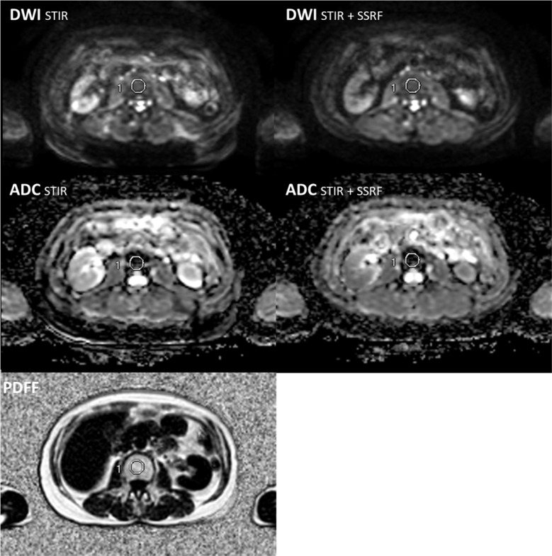
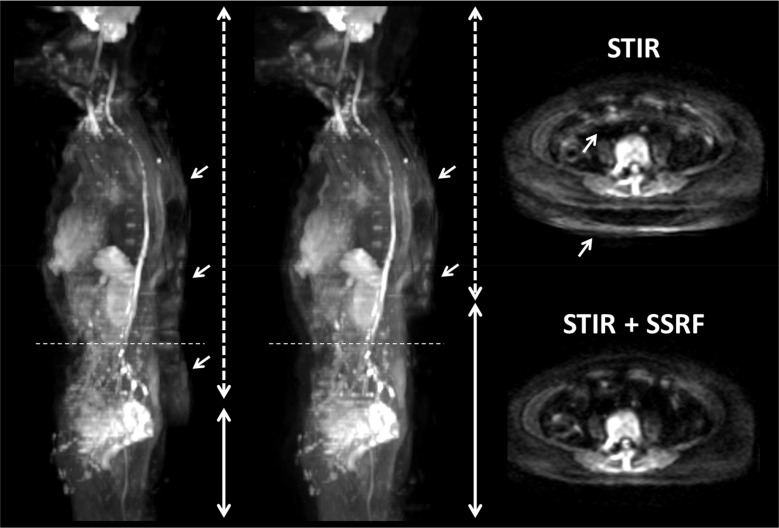
Seventy-seven patients underwent whole-body DWI with short-inversion time inversion-recovery (STIR) (DWI) and/or STIR + selective water-excitation (spectral-spatial RF [SSRF]) (DWI). ADCs of lumbar vertebrae (L3 and L4) were compared between DWI and DWI, and correlated with the PDFF.
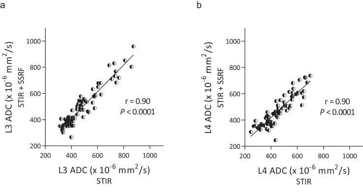
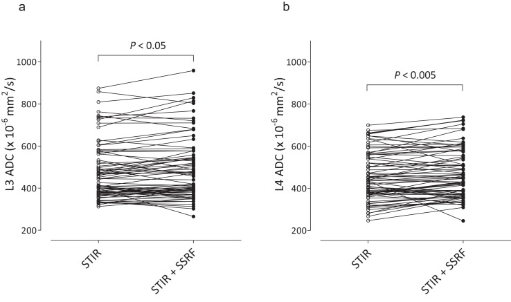
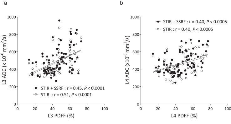
Lumbar ADCs obtained by DWI and DWI were significantly correlated (L3: r = 0.90, P < 0.0001, L4: r = 0.90, P < 0.0001). Lumbar ADCs (× 10 mm/s) obtained by DWI were significantly lower than those by DWI (L3: 479 ± 137 and 490 ± 148, P < 0.05, L4: 456 ± 114 and 471 ± 118, P < 0.005). Residual fat signals were more clearly observed on DWI than on DWI. The ADCs of L3 obtained by DWI and DWI exhibited significant positive correlations with the PDFF (r = 0.51, P < 0.0001, and r = 0.45, P < 0.0001, respectively), and the ADCs of L4 obtained by DWI and DWI exhibited significantly positive correlations with the PDFF (r = 0.40, P < 0.0005, and r = 0.40, P < 0.0005, respectively).
Irrespective of different fat-suppression methods, lumbar ADCs were positively correlated with the PDFF, being inconsistent with previous studies. Lumbar ADCs obtained by DWI were significantly lower than those obtained by DWI probably due to residual fat signals on DWI. However, this difference (< 4%) did not explain the positive correlation between lumbar ADC and PDFF.
比较两种脂肪抑制技术在弥散加权成像(DWI)中骨髓的表观弥散系数(ADC),并评估骨髓 ADC 与质子密度脂肪分数(PDFF)之间的相关性。
77 例患者行全身 DWI 检查,采用短反转时间反转恢复(STIR)(DWI)和/或 STIR+选择性水激发(谱空间射频[SSRF])(DWI)。比较 L3 和 L4 腰椎的 DWI 和 DWI 的 ADC 值,并与 PDFF 相关。
DWI 和 DWI 获得的腰椎 ADC 值呈显著相关(L3:r=0.90,P<0.0001,L4:r=0.90,P<0.0001)。DWI 获得的腰椎 ADC 值(×10mm/s)明显低于 DWI(L3:479±137 和 490±148,P<0.05,L4:456±114 和 471±118,P<0.005)。DWI 上残留的脂肪信号比 DWI 上更清晰。DWI 和 DWI 获得的 L3 ADC 值与 PDFF 呈显著正相关(r=0.51,P<0.0001 和 r=0.45,P<0.0001),DWI 和 DWI 获得的 L4 ADC 值与 PDFF 呈显著正相关(r=0.40,P<0.0005 和 r=0.40,P<0.0005)。
无论脂肪抑制方法如何,腰椎 ADC 值与 PDFF 呈正相关,与以往的研究结果不一致。DWI 获得的腰椎 ADC 值明显低于 DWI 获得的 ADC 值,可能是因为 DWI 上有残留的脂肪信号。然而,这种差异(<4%)并不能解释腰椎 ADC 与 PDFF 之间的正相关关系。