Department of Veterinary Physiology and Biochemistry, Justus-Liebig-University Giessen, Frankfurter Strasse 100, 35392, Giessen, Germany.
Inflamm Res. 2021 Apr;70(4):429-444. doi: 10.1007/s00011-021-01440-7. Epub 2021 Feb 13.
Bacterial lipopolysaccharide (LPS) may contribute to the manifestation of inflammatory pain within structures of the afferent somatosensory system. LPS can induce a state of refractoriness to its own effects termed LPS tolerance. We employed primary neuro-glial cultures from rat dorsal root ganglia (DRG) and the superficial dorsal horn (SDH) of the spinal cord, mainly including the substantia gelatinosa to establish and characterize a model of LPS tolerance within these structures.
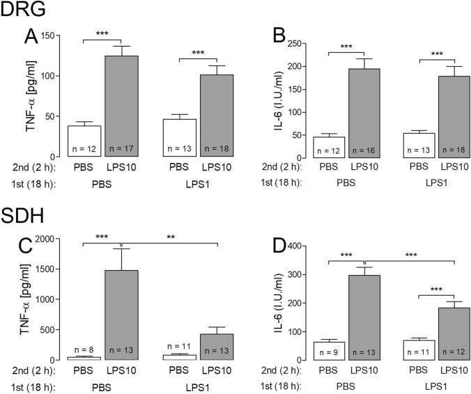
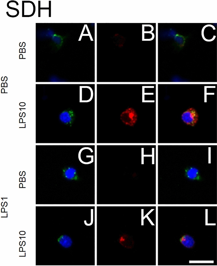
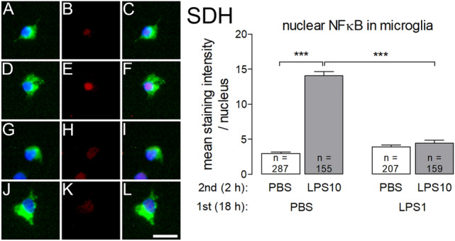
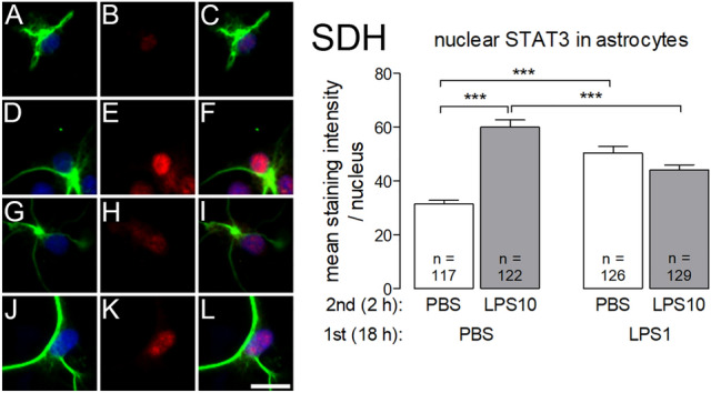
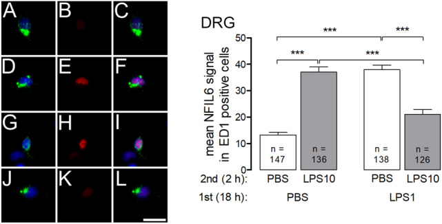
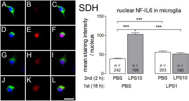
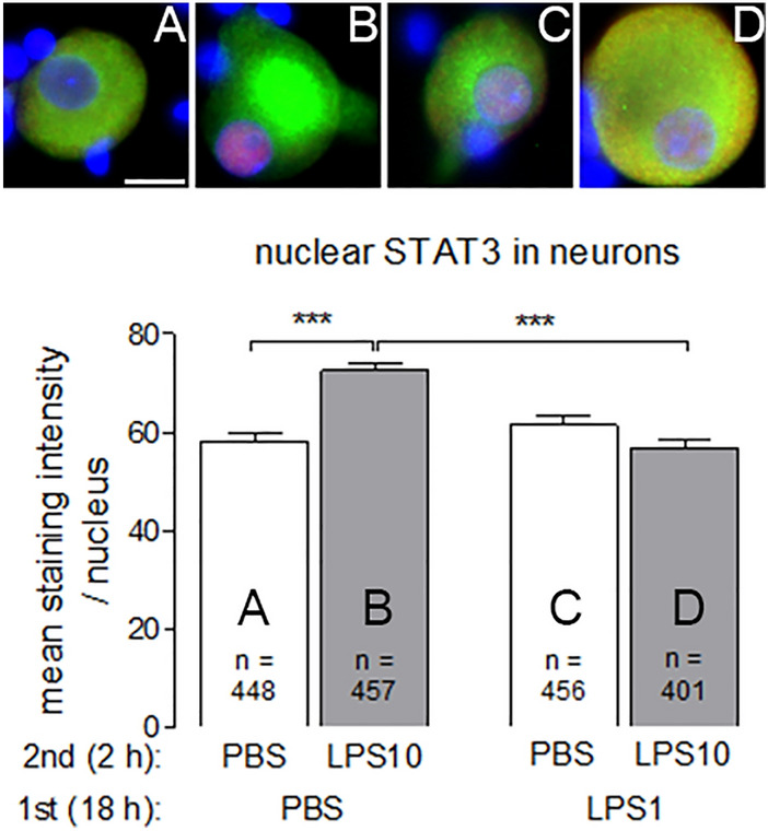
Tolerance was induced by pre-treatment of both cultures with 1 µg/ml LPS for 18 h, followed by a short-term stimulation with a higher LPS dose (10 µg/ml for 2 h). Cultures treated with solvent were used as controls. Cells from DRG or SDH were investigated by means of RT-PCR (expression of inflammatory genes) and immunocytochemistry (translocation of inflammatory transcription factors into nuclei of cells from both cultures). Supernatants from both cultures were assayed for tumor necrosis factor-α (TNF-α) and interleukin-6 (IL-6) by highly sensitive bioassays.
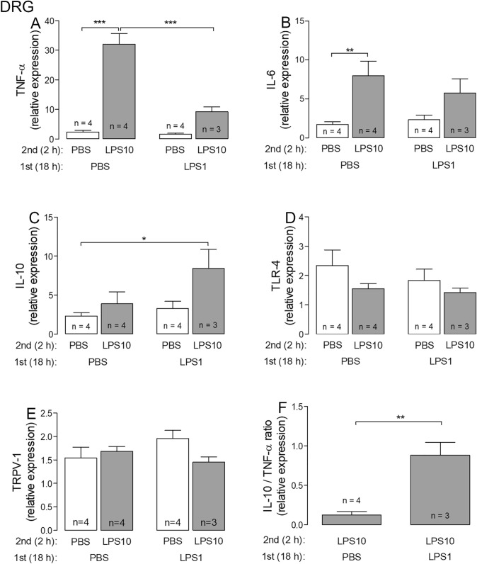
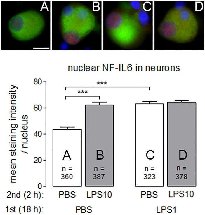
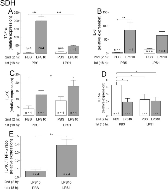
At the mRNA-level, pre-treatment with 1 µg/ml LPS caused reduced expression of TNF-α and enhanced IL-10/TNF-α expression ratios in both cultures upon subsequent stimulation with 10 µg/ml LPS, i.e. LPS tolerance. SDH cultures further showed reduced release of TNF-α into the supernatants and attenuated TNF-α immunoreactivity in microglial cells. In the state of LPS tolerance macrophages from DRG and microglial cells from SDH showed reduced LPS-induced nuclear translocation of the inflammatory transcription factors NFκB and NF-IL6. Nuclear immunoreactivity of the IL-6-activated transcription factor STAT3 was further reduced in neurons from DRG and astrocytes from SDH in LPS tolerant cultures.
A state of LPS tolerance can be induced in primary cultures from the afferent somatosensory system, which is characterized by a down-regulation of pro-inflammatory mediators. Thus, this model can be applied to study the effects of LPS tolerance at the cellular level, for example possible modifications of neuronal reactivity patterns upon inflammatory stimulation.
细菌脂多糖(LPS)可能会导致传入感觉系统结构中炎症性疼痛的表现。LPS 可以诱导对自身效应的不应答状态,称为 LPS 耐受。我们采用大鼠背根神经节(DRG)和脊髓浅层背角(SDH)的原代神经胶质培养物,主要包括胶状质,在这些结构中建立并表征 LPS 耐受模型。
通过用 1μg/ml LPS 预处理两种培养物 18 小时来诱导耐受,然后用较高 LPS 剂量(10μg/ml 持续 2 小时)进行短期刺激。用溶剂处理的培养物作为对照。通过 RT-PCR(炎症基因的表达)和免疫细胞化学(两种培养物的细胞中炎症转录因子的易位)研究 DRG 或 SDH 中的细胞。通过高灵敏度生物测定法测定两种培养物上清液中的肿瘤坏死因子-α(TNF-α)和白细胞介素-6(IL-6)。
在 mRNA 水平上,用 1μg/ml LPS 预处理导致两种培养物在用 10μg/ml LPS 随后刺激时 TNF-α 的表达减少,并增强了 IL-10/TNF-α 的表达比率,即 LPS 耐受。SDH 培养物进一步显示 TNF-α 向上清液中的释放减少,并且 TNF-α 免疫反应性在小胶质细胞中减弱。在 LPS 耐受状态下,DRG 中的巨噬细胞和 SDH 中的小胶质细胞表现出 LPS 诱导的炎症转录因子 NFκB 和 NF-IL6 的核易位减少。在 LPS 耐受培养物中,神经元中的 IL-6 激活转录因子 STAT3 的核免疫反应性进一步降低,SDH 中的星形胶质细胞。
可以在传入感觉系统的原代培养物中诱导 LPS 耐受状态,其特征在于促炎介质的下调。因此,该模型可用于在细胞水平上研究 LPS 耐受的影响,例如炎症刺激下神经元反应模式的可能改变。