Bhuva Anish N, Treibel Thomas A, Seraphim Andreas, Scully Paul, Knott Kristopher D, Augusto João B, Torlasco Camilla, Menacho Katia, Lau Clement, Patel Kush, Moon James C, Kellman Peter, Manisty Charlotte H
Institute for Cardiovascular Science, University College London, London, United Kingdom.
Department of Cardiovascular Imaging, Barts Heart Centre, Barts Health NHS Trust, London, United Kingdom.
Front Cardiovasc Med. 2021 Jan 29;8:631366. doi: 10.3389/fcvm.2021.631366. eCollection 2021.
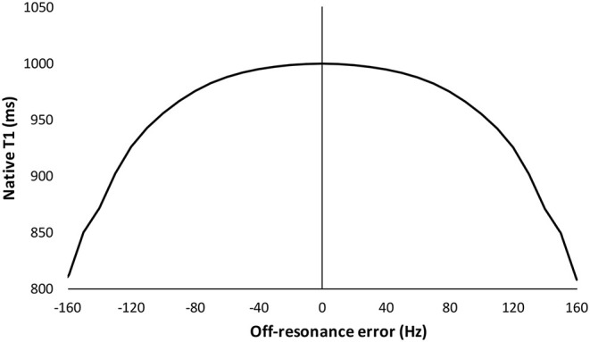
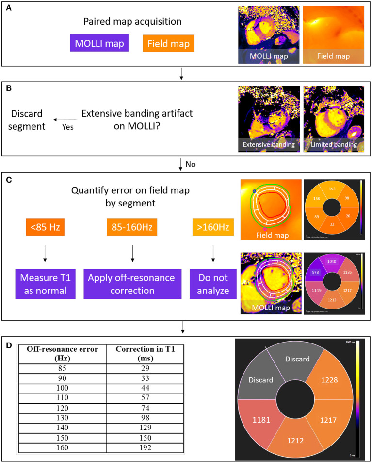
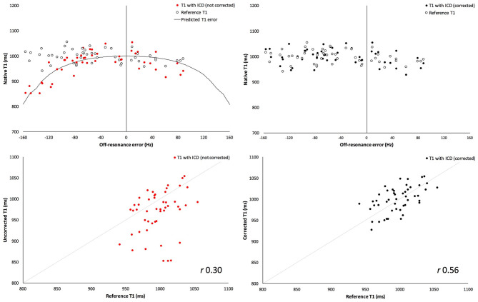
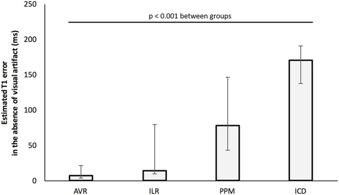
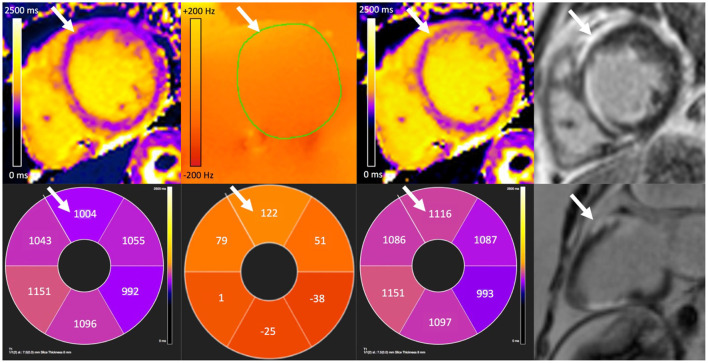
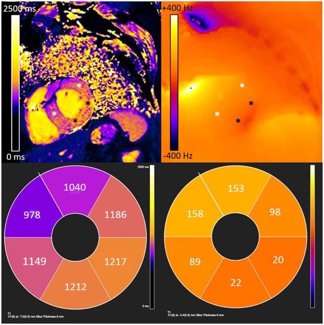
Measurement of myocardial T1 is increasingly incorporated into standard cardiovascular magnetic resonance (CMR) protocols, however accuracy may be reduced in patients with metallic cardiovascular implants. Measurement is feasible in segments free from visual artifact, but there may still be off-resonance induced error. To quantify off-resonance induced T1 error in patients with metallic cardiovascular implants, and validate a method for error correction for a conventional MOLLI pulse sequence. Twenty-four patients with cardiac implantable electronic devices (CIEDs: 46% permanent pacemakers, PPMs; 33% implantable loop recorders, ILRs; and 21% implantable cardioverter-defibrillators, ICDs); and 31 patients with aortic valve replacement (AVR) (45% metallic) were studied. Paired mid-myocardial short-axis MOLLI and single breath-hold off-resonance field maps were acquired at 1.5 T. T1 values were measured by AHA segment, and segments with visual artifact were excluded. T1 correction was applied using a published relationship between off-resonance and T1. The accuracy of the correction was assessed in 10 healthy volunteers by measuring T1 before and after external placement of an ICD generator next to the chest to generate off-resonance. T1 values in healthy volunteers with an ICD were underestimated compared to without (967 ± 52 vs. 997 ± 26 ms respectively, = 0.0001), but were similar after correction ( = 0.57, residual difference 2 ± 27 ms). Artifact was visible in 4 ± 12, 42 ± 31, and 53 ± 27% of AHA segments in patients with ILRs, PPMs, and ICDs, respectively. In segments without artifact, T1 was underestimated by 63 ms (interquartile range: 7-143) per patient. The greatest error for patients with ILRs, PPMs and ICDs were 79, 146, and 191 ms, respectively. The presence of an AVR did not generate T1 error. Even when there is no visual artifact, there is error in T1 in patients with CIEDs, but not AVRs. Off-resonance field map acquisition can detect error in measured T1, and a correction can be applied to quantify T1 MOLLI accurately.
心肌T1测量越来越多地被纳入标准心血管磁共振(CMR)检查方案中,然而,对于有金属心血管植入物的患者,测量准确性可能会降低。在无视觉伪影的节段中测量是可行的,但仍可能存在失谐诱导误差。为了量化有金属心血管植入物患者中失谐诱导的T1误差,并验证一种针对传统MOLLI脉冲序列的误差校正方法。对24例植入心脏电子设备的患者(心脏植入式电子设备:46%为永久性起搏器,PPM;33%为植入式循环记录仪,ILR;21%为植入式心脏复律除颤器,ICD)以及31例接受主动脉瓣置换术(AVR)的患者(45%为金属瓣膜)进行了研究。在1.5T条件下采集配对的心肌中层短轴MOLLI图像和单次屏气失谐场图。按美国心脏协会(AHA)节段测量T1值,排除有视觉伪影的节段。利用已发表的失谐与T1之间的关系进行T1校正。通过在10名健康志愿者胸部旁边外置ICD发生器以产生失谐,测量外置前后的T1值,评估校正的准确性。与未外置ICD的健康志愿者相比,有ICD的健康志愿者的T1值被低估(分别为967±52ms和997±26ms,P=0.0001),但校正后相似(P=0.57,残余差异为2±27ms)。ILR、PPM和ICD患者中,分别有4±12%、42±31%和53±27%的AHA节段可见伪影。在无伪影的节段中,每位患者的T1被低估63ms(四分位间距:7 - 143)。ILR、PPM和ICD患者的最大误差分别为79、146和191ms。AVR的存在未产生T1误差。即使没有视觉伪影,有心脏植入式电子设备的患者的T1也存在误差,但接受主动脉瓣置换术的患者不存在。采集失谐场图可检测测量T1中的误差,并且可以应用校正来准确量化T1 MOLLI值。