• 文献检索
  • 文档翻译
  • 深度研究
  • 学术资讯
  • Suppr Zotero 插件Zotero 插件
  • 邀请有礼
  • 套餐&价格
  • 历史记录
应用&插件
Suppr Zotero 插件Zotero 插件浏览器插件Mac 客户端Windows 客户端微信小程序
定价
高级版会员购买积分包购买API积分包
服务
文献检索文档翻译深度研究API 文档MCP 服务
关于我们
关于 Suppr公司介绍联系我们用户协议隐私条款
关注我们

Suppr 超能文献

核心技术专利:CN118964589B侵权必究
粤ICP备2023148730 号-1Suppr @ 2026

文献检索

告别复杂PubMed语法,用中文像聊天一样搜索,搜遍4000万医学文献。AI智能推荐,让科研检索更轻松。

立即免费搜索

文件翻译

保留排版,准确专业,支持PDF/Word/PPT等文件格式,支持 12+语言互译。

免费翻译文档

深度研究

AI帮你快速写综述,25分钟生成高质量综述,智能提取关键信息,辅助科研写作。

立即免费体验

深度学习算法在胸部 X 线片上对可转诊的胸部异常的性能:一项健康筛查队列的多中心研究。

Performance of a deep-learning algorithm for referable thoracic abnormalities on chest radiographs: A multicenter study of a health screening cohort.

机构信息

Department of Radiology, Gil Medical Center, Gachon University College of Medicine, Incheon, South Korea.

Department of Biomedical Engineering, Gachon University College of Medicine, Incheon, South Korea.

出版信息

PLoS One. 2021 Feb 19;16(2):e0246472. doi: 10.1371/journal.pone.0246472. eCollection 2021.

DOI:10.1371/journal.pone.0246472
PMID:33606779
原文链接:https://pmc.ncbi.nlm.nih.gov/articles/PMC7894861/
Abstract

PURPOSE

This study evaluated the performance of a commercially available deep-learning algorithm (DLA) (Insight CXR, Lunit, Seoul, South Korea) for referable thoracic abnormalities on chest X-ray (CXR) using a consecutively collected multicenter health screening cohort.

METHODS AND MATERIALS

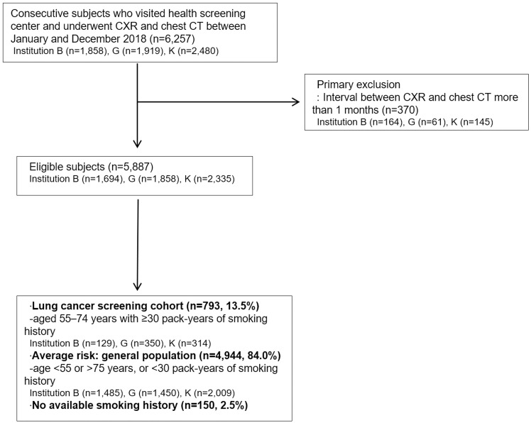
A consecutive health screening cohort of participants who underwent both CXR and chest computed tomography (CT) within 1 month was retrospectively collected from three institutions' health care clinics (n = 5,887). Referable thoracic abnormalities were defined as any radiologic findings requiring further diagnostic evaluation or management, including DLA-target lesions of nodule/mass, consolidation, or pneumothorax. We evaluated the diagnostic performance of the DLA for referable thoracic abnormalities using the area under the receiver operating characteristic (ROC) curve (AUC), sensitivity, and specificity using ground truth based on chest CT (CT-GT). In addition, for CT-GT-positive cases, three independent radiologist readings were performed on CXR and clear visible (when more than two radiologists called) and visible (at least one radiologist called) abnormalities were defined as CXR-GTs (clear visible CXR-GT and visible CXR-GT, respectively) to evaluate the performance of the DLA.

RESULTS

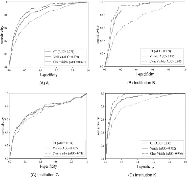
Among 5,887 subjects (4,329 males; mean age 54±11 years), referable thoracic abnormalities were found in 618 (10.5%) based on CT-GT. DLA-target lesions were observed in 223 (4.0%), nodule/mass in 202 (3.4%), consolidation in 31 (0.5%), pneumothorax in one 1 (<0.1%), and DLA-non-target lesions in 409 (6.9%). For referable thoracic abnormalities based on CT-GT, the DLA showed an AUC of 0.771 (95% confidence interval [CI], 0.751-0.791), a sensitivity of 69.6%, and a specificity of 74.0%. Based on CXR-GT, the prevalence of referable thoracic abnormalities decreased, with visible and clear visible abnormalities found in 405 (6.9%) and 227 (3.9%) cases, respectively. The performance of the DLA increased significantly when using CXR-GTs, with an AUC of 0.839 (95% CI, 0.829-0.848), a sensitivity of 82.7%, and s specificity of 73.2% based on visible CXR-GT and an AUC of 0.872 (95% CI, 0.863-0.880, P <0.001 for the AUC comparison of GT-CT vs. clear visible CXR-GT), a sensitivity of 83.3%, and a specificity of 78.8% based on clear visible CXR-GT.

CONCLUSION

The DLA provided fair-to-good stand-alone performance for the detection of referable thoracic abnormalities in a multicenter consecutive health screening cohort. The DLA showed varied performance according to the different methods of ground truth.

摘要

目的

本研究使用连续采集的多中心健康筛查队列,评估一种商用深度学习算法(DLA)(Insight CXR,Lunit,韩国首尔)在胸部 X 线(CXR)上检测有意义的胸部异常的性能。

方法和材料

从三个机构的医疗诊所回顾性收集了一个连续的健康筛查队列,其中参与者在 1 个月内同时接受了 CXR 和胸部计算机断层扫描(CT)(n=5887)。有意义的胸部异常定义为任何需要进一步诊断评估或管理的影像学发现,包括 DLA 目标病变(结节/肿块、实变或气胸)。我们使用基于胸部 CT 的地面实况(CT-GT)评估了 DLA 对有意义的胸部异常的诊断性能,使用受试者工作特征(ROC)曲线下面积(AUC)、敏感性和特异性。此外,对于 CT-GT 阳性病例,对 CXR 进行了三名独立放射科医生的阅读,将明确可见(当两名以上放射科医生称有异常时)和可见(至少有一名放射科医生称有异常)的异常定义为 CXR-GT(明确可见 CXR-GT 和可见 CXR-GT),以评估 DLA 的性能。

结果

在 5887 名受试者中(4329 名男性;平均年龄 54±11 岁),根据 CT-GT 发现 618 名(10.5%)有意义的胸部异常。DLA 目标病变见于 223 例(4.0%),结节/肿块见于 202 例(3.4%),实变见于 31 例(0.5%),气胸见于 1 例(<0.1%),DLA 非目标病变见于 409 例(6.9%)。对于基于 CT-GT 的有意义的胸部异常,DLA 的 AUC 为 0.771(95%置信区间,0.751-0.791),敏感性为 69.6%,特异性为 74.0%。基于 CXR-GT,有意义的胸部异常的患病率降低,可见异常和明确可见异常分别见于 405 例(6.9%)和 227 例(3.9%)。当使用 CXR-GT 时,DLA 的性能显著提高,可见 CXR-GT 的 AUC 为 0.839(95%置信区间,0.829-0.848),敏感性为 82.7%,特异性为 73.2%,明确可见 CXR-GT 的 AUC 为 0.872(95%置信区间,0.863-0.880,P<0.001 用于 GT-CT 与明确可见 CXR-GT 的 AUC 比较),敏感性为 83.3%,特异性为 78.8%。

结论

DLA 在多中心连续健康筛查队列中对有意义的胸部异常的检测提供了良好的独立性能。DLA 根据不同的地面实况方法表现出不同的性能。

https://cdn.ncbi.nlm.nih.gov/pmc/blobs/bd1f/7894861/0f2af525c570/pone.0246472.g003.jpg
https://cdn.ncbi.nlm.nih.gov/pmc/blobs/bd1f/7894861/8d4f4c022240/pone.0246472.g001.jpg
https://cdn.ncbi.nlm.nih.gov/pmc/blobs/bd1f/7894861/7ec118d8c7e3/pone.0246472.g002.jpg
https://cdn.ncbi.nlm.nih.gov/pmc/blobs/bd1f/7894861/0f2af525c570/pone.0246472.g003.jpg
https://cdn.ncbi.nlm.nih.gov/pmc/blobs/bd1f/7894861/8d4f4c022240/pone.0246472.g001.jpg
https://cdn.ncbi.nlm.nih.gov/pmc/blobs/bd1f/7894861/7ec118d8c7e3/pone.0246472.g002.jpg
https://cdn.ncbi.nlm.nih.gov/pmc/blobs/bd1f/7894861/0f2af525c570/pone.0246472.g003.jpg

相似文献

1
Performance of a deep-learning algorithm for referable thoracic abnormalities on chest radiographs: A multicenter study of a health screening cohort.深度学习算法在胸部 X 线片上对可转诊的胸部异常的性能:一项健康筛查队列的多中心研究。
PLoS One. 2021 Feb 19;16(2):e0246472. doi: 10.1371/journal.pone.0246472. eCollection 2021.
2
Concordance rate of radiologists and a commercialized deep-learning solution for chest X-ray: Real-world experience with a multicenter health screening cohort.放射科医生与商业化深度学习解决方案在胸部 X 光片检测中的符合率:多中心健康筛查队列的真实世界经验。
PLoS One. 2022 Feb 24;17(2):e0264383. doi: 10.1371/journal.pone.0264383. eCollection 2022.
3
Utility of a Deep Learning Algorithm for Detection of Reticular Opacity on Chest Radiography in Patients With Interstitial Lung Disease.深度学习算法在间质性肺疾病患者胸部 X 线摄影中检测网状混浊的效用。
AJR Am J Roentgenol. 2022 Apr;218(4):642-650. doi: 10.2214/AJR.21.26682. Epub 2021 Oct 20.
4
Deep learning in chest radiography: Detection of findings and presence of change.深度学习在胸部 X 光摄影中的应用:检测结果和变化的存在。
PLoS One. 2018 Oct 4;13(10):e0204155. doi: 10.1371/journal.pone.0204155. eCollection 2018.
5
Diagnostic effect of artificial intelligence solution for referable thoracic abnormalities on chest radiography: a multicenter respiratory outpatient diagnostic cohort study.人工智能解决方案对胸部放射摄影中可转诊胸部异常的诊断效果:一项多中心呼吸门诊诊断队列研究。
Eur Radiol. 2022 May;32(5):3469-3479. doi: 10.1007/s00330-021-08397-5. Epub 2022 Jan 1.
6
Artificial Intelligence-Based Identification of Normal Chest Radiographs: A Simulation Study in a Multicenter Health Screening Cohort.基于人工智能的正常胸部 X 光片识别:多中心健康筛查队列中的模拟研究。
Korean J Radiol. 2022 Oct;23(10):1009-1018. doi: 10.3348/kjr.2022.0189.
7
Performance of a Deep Learning Algorithm Compared with Radiologic Interpretation for Lung Cancer Detection on Chest Radiographs in a Health Screening Population.深度学习算法与放射解读在健康筛查人群中对胸部 X 光片肺癌检测的性能比较。
Radiology. 2020 Dec;297(3):687-696. doi: 10.1148/radiol.2020201240. Epub 2020 Sep 22.
8
Deep learning algorithm for surveillance of pneumothorax after lung biopsy: a multicenter diagnostic cohort study.深度学习算法在肺活检后气胸监测中的应用:一项多中心诊断队列研究。
Eur Radiol. 2020 Jul;30(7):3660-3671. doi: 10.1007/s00330-020-06771-3. Epub 2020 Mar 11.
9
Using AI to Improve Radiologist Performance in Detection of Abnormalities on Chest Radiographs.利用人工智能提高放射科医生在胸部X光片上检测异常的表现。
Radiology. 2023 Dec;309(3):e230860. doi: 10.1148/radiol.230860.
10
Comparison of Baseline, Bone-Subtracted, and Enhanced Chest Radiographs for Detection of Pneumothorax.比较基线、骨减去和增强胸部 X 线片在气胸检测中的表现。
Can Assoc Radiol J. 2021 Aug;72(3):519-524. doi: 10.1177/0846537120908852. Epub 2020 Mar 18.

引用本文的文献

1
Factors for increasing positive predictive value of pneumothorax detection on chest radiographs using artificial intelligence.利用人工智能提高胸部 X 光片气胸检测阳性预测值的因素。
Sci Rep. 2024 Aug 23;14(1):19624. doi: 10.1038/s41598-024-70780-1.
2
Artificial Intelligence-Based Software with CE Mark for Chest X-ray Interpretation: Opportunities and Challenges.具有CE标志的基于人工智能的胸部X光解读软件:机遇与挑战。
Diagnostics (Basel). 2023 Jun 10;13(12):2020. doi: 10.3390/diagnostics13122020.
3
Deep learning for pneumothorax diagnosis: a systematic review and meta-analysis.

本文引用的文献

1
Deep Learning Based Lung Region Segmentation with Data Preprocessing by Generative Adversarial Nets.基于深度学习的肺部区域分割与生成对抗网络的数据预处理
Annu Int Conf IEEE Eng Med Biol Soc. 2020 Jul;2020:1278-1281. doi: 10.1109/EMBC44109.2020.9176214.
2
Lung Region Segmentation in Chest X-Ray Images using Deep Convolutional Neural Networks.使用深度卷积神经网络对胸部X光图像中的肺部区域进行分割
Annu Int Conf IEEE Eng Med Biol Soc. 2020 Jul;2020:1246-1249. doi: 10.1109/EMBC44109.2020.9175478.
3
MIMIC-CXR, a de-identified publicly available database of chest radiographs with free-text reports.
深度学习在气胸诊断中的应用:系统评价和荟萃分析。
Eur Respir Rev. 2023 Jun 7;32(168). doi: 10.1183/16000617.0259-2022. Print 2023 Jun 30.
4
The rise of artificial intelligence reading of chest X-rays for enhanced TB diagnosis and elimination.人工智能读片在增强结核病诊断和消除中的应用。
Int J Tuberc Lung Dis. 2023 May 1;27(5):367-372. doi: 10.5588/ijtld.22.0687.
5
Hospital-wide survey of clinical experience with artificial intelligence applied to daily chest radiographs.全院范围内对人工智能在日常胸部 X 光片中应用的临床经验调查。
PLoS One. 2023 Mar 2;18(3):e0282123. doi: 10.1371/journal.pone.0282123. eCollection 2023.
6
Artificial Intelligence-Based Identification of Normal Chest Radiographs: A Simulation Study in a Multicenter Health Screening Cohort.基于人工智能的正常胸部 X 光片识别:多中心健康筛查队列中的模拟研究。
Korean J Radiol. 2022 Oct;23(10):1009-1018. doi: 10.3348/kjr.2022.0189.
7
Localization-adjusted diagnostic performance and assistance effect of a computer-aided detection system for pneumothorax and consolidation.气胸和实变计算机辅助检测系统的定位调整诊断性能及辅助效果
NPJ Digit Med. 2022 Jul 30;5(1):107. doi: 10.1038/s41746-022-00658-x.
8
Development and Validation of a Multimodal-Based Prognosis and Intervention Prediction Model for COVID-19 Patients in a Multicenter Cohort.基于多模态的多中心队列 COVID-19 患者预后和干预预测模型的建立和验证。
Sensors (Basel). 2022 Jul 2;22(13):5007. doi: 10.3390/s22135007.
9
Concordance rate of radiologists and a commercialized deep-learning solution for chest X-ray: Real-world experience with a multicenter health screening cohort.放射科医生与商业化深度学习解决方案在胸部 X 光片检测中的符合率:多中心健康筛查队列的真实世界经验。
PLoS One. 2022 Feb 24;17(2):e0264383. doi: 10.1371/journal.pone.0264383. eCollection 2022.
10
Correction: Performance of a deep-learning algorithm for referable thoracic abnormalities on chest radiographs: A multicenter study of a health screening cohort.更正:用于胸部X线片上可参考的胸部异常的深度学习算法的性能:一项健康筛查队列的多中心研究。
PLoS One. 2021 Apr 28;16(4):e0251045. doi: 10.1371/journal.pone.0251045. eCollection 2021.
MIMIC-CXR,一个去标识化的、公开可用的、包含自由文本报告的胸部 X 光数据库。
Sci Data. 2019 Dec 12;6(1):317. doi: 10.1038/s41597-019-0322-0.
4
Development and Validation of a Deep Learning-Based Automated Detection Algorithm for Major Thoracic Diseases on Chest Radiographs.基于深度学习的胸部 X 线片主要胸部疾病自动检测算法的开发与验证。
JAMA Netw Open. 2019 Mar 1;2(3):e191095. doi: 10.1001/jamanetworkopen.2019.1095.
5
Deep learning for chest radiograph diagnosis: A retrospective comparison of the CheXNeXt algorithm to practicing radiologists.深度学习在胸片诊断中的应用:CheXNeXt 算法与临床放射科医生的回顾性比较。
PLoS Med. 2018 Nov 20;15(11):e1002686. doi: 10.1371/journal.pmed.1002686. eCollection 2018 Nov.
6
Development and Validation of a Deep Learning-based Automatic Detection Algorithm for Active Pulmonary Tuberculosis on Chest Radiographs.基于深度学习的胸部 X 线片活动性肺结核自动检测算法的开发与验证。
Clin Infect Dis. 2019 Aug 16;69(5):739-747. doi: 10.1093/cid/ciy967.
7
Development and Validation of Deep Learning-based Automatic Detection Algorithm for Malignant Pulmonary Nodules on Chest Radiographs.基于深度学习的胸部 X 线片恶性肺结节自动检测算法的开发与验证。
Radiology. 2019 Jan;290(1):218-228. doi: 10.1148/radiol.2018180237. Epub 2018 Sep 25.
8
Korean National Health Insurance Database.韩国国民健康保险数据库。
JAMA Intern Med. 2016 Jan;176(1):138. doi: 10.1001/jamainternmed.2015.7110.
9
Common patterns in 558 diagnostic radiology errors.558例诊断性放射学错误的常见模式。
J Med Imaging Radiat Oncol. 2012 Apr;56(2):173-8. doi: 10.1111/j.1754-9485.2012.02348.x.
10
Fleischner Society: glossary of terms for thoracic imaging.弗莱施纳学会:胸部影像学术语词汇表。
Radiology. 2008 Mar;246(3):697-722. doi: 10.1148/radiol.2462070712. Epub 2008 Jan 14.