Department of Chemistry, KAIST, Daejeon, Korea.
Cannabis Cannabinoid Res. 2021 Feb 12;6(1):40-47. doi: 10.1089/can.2019.0102. eCollection 2021.
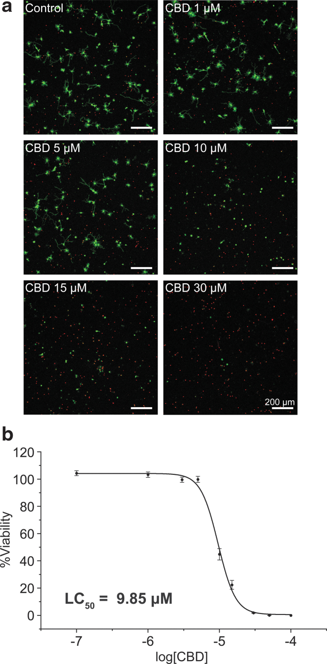
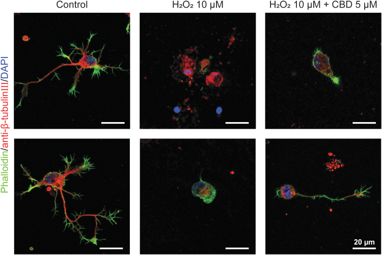
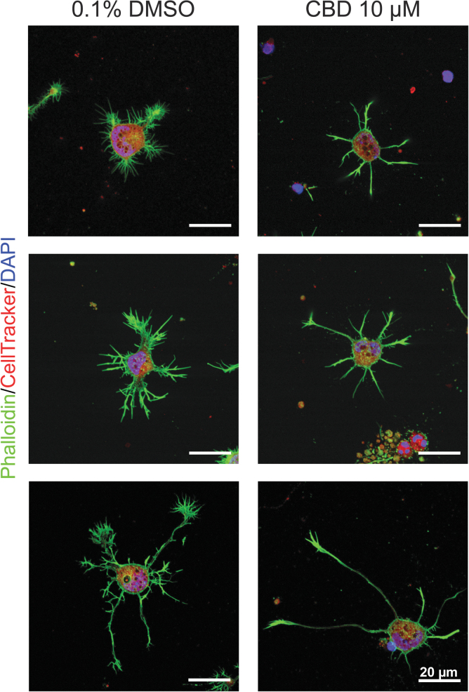
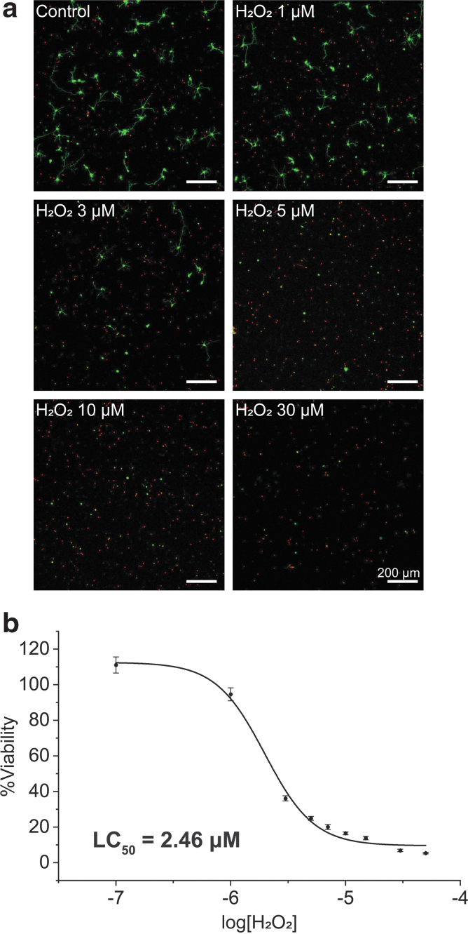
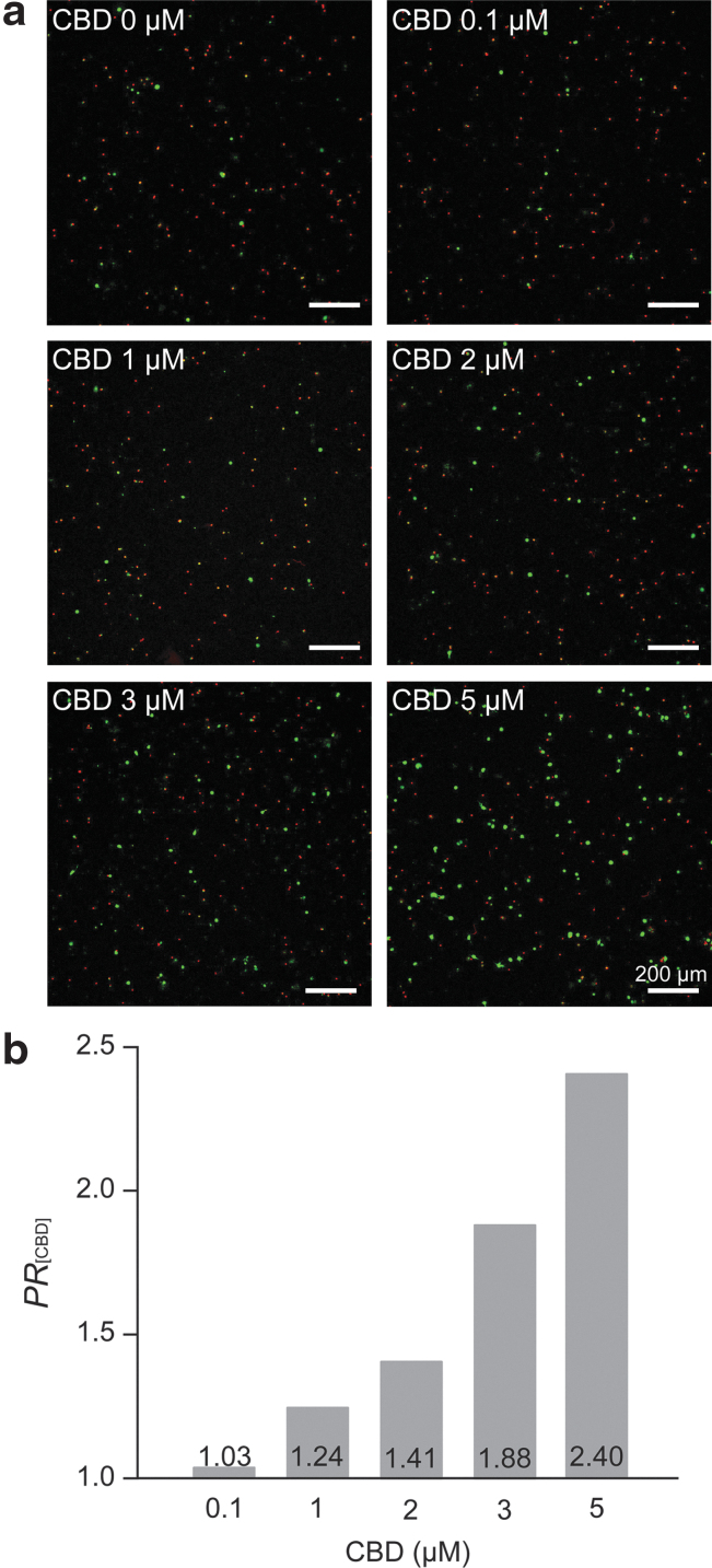
Reports on the neurotoxic and neuroprotective effects of cannabidiol (CBD) have not been in complete accord, showing different and somewhat contradictory results depending upon the brain cell types and experimental conditions employed. This work systematically examines the neuroprotective capability of CBD against oxidative stress (i.e., hydrogen peroxide [HO]) as well as its toxicity profile in the culture platform of primary hippocampal neurons. The low cell-density (100 neurons per mm) culture was used for analyzing the viability and morphology of neurons at a single-cell level with a confocal laser-scanning microscope (CLSM). Primary neurons were obtained from the hippocampal tissues of embryonic day-18 (E18) Sprague-Dawley rat pups and treated with CBD (0.1-100 μM) and/or HO (0.1-50 μM) at 1 DIV (days ). The lethal concentration 50 (LC) value (the concentration causing 50% cell death) of CBD was calculated to be 9.85 μM after 24 h of incubation, and that of HO was 2.46 μM under the same conditions. The neuroprotection ratio of CBD against HO ([HO]=10 μM) was 2.40 with 5 μM of CBD, increasing the cell viability to 57% from 24%. The CLSM analysis suggested that the cell-death mechanisms were different for CBD and HO, and CBD did not completely rescue the morphological alterations of primary hippocampal neurons caused by HO, such as neurite degeneration, at least in the neuron culture. Although CBD showed both neurotoxic and neuroprotective effects on hippocampal neurons in the setting, the use of low-concentrated (i.e., 5 μM) CBD, not causing toxic effects on the neurons, significantly rescued the neurons from the oxidative stress (HO), confirming its neuroprotection capability.
关于大麻二酚(CBD)的神经毒性和神经保护作用的报告并不完全一致,根据所使用的脑细胞类型和实验条件,显示出不同的、有些矛盾的结果。本工作系统地研究了 CBD 对抗氧化应激(即过氧化氢[HO])的神经保护能力及其在原代海马神经元培养物中的毒性谱。低细胞密度(100 个神经元/平方毫米)培养物用于在单细胞水平上用共聚焦激光扫描显微镜(CLSM)分析神经元的活力和形态。原代神经元从胚胎期 18 天(E18)的 Sprague-Dawley 大鼠海马组织中获得,并在 1 天分裂(DIV)时用 CBD(0.1-100 μM)和/或 HO(0.1-50 μM)处理。孵育 24 小时后,CBD 的致死浓度 50(LC)值(引起 50%细胞死亡的浓度)为 9.85 μM,HO 的 LC 值为 2.46 μM。在相同条件下,CBD 对 HO 的神经保护比为 2.40,用 5 μM 的 CBD,将细胞活力从 24%提高到 57%。CLSM 分析表明,CBD 和 HO 的细胞死亡机制不同,CBD 并不能完全挽救 HO 引起的原代海马神经元的形态改变,例如轴突退化,至少在神经元培养物中是这样。虽然 CBD 在这种情况下对海马神经元表现出神经毒性和神经保护作用,但使用低浓度(即 5 μM)的 CBD 不会对神经元产生毒性作用,能显著挽救神经元免受氧化应激(HO)的影响,证实了其神经保护能力。