Masumoto Akiko, Kitai Takeshi, Ota Mitsuhiko, Kim Kitae, Ehara Natsuhiko, Furukawa Yutaka
Department of Cardiovascular Medicine, Kobe City Medical Center General Hospital, 2-1-1 Minatojima-minamimachi, Chuo-ku, Kobe 6500047, Japan.
Eur Heart J Case Rep. 2020 Dec 7;4(6):1-4. doi: 10.1093/ehjcr/ytaa392. eCollection 2020 Dec.
Increasing number of symptomatic patients with severe aortic stenosis is treated with transcatheter aortic valve implantation (TAVI). Stroke is one of the most serious complications of TAVI, and the majority of cerebral events in patients undergoing TAVI have an embolic origin.
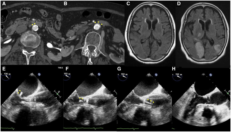
A 90-year-old female underwent trans-femoral TAVI for symptomatic severe aortic stenosis. Just before the implantation of the transcatheter heart valve (THV), transoesophageal echocardiography (TOE) showed a mobile, high-echoic mass attached to the THV, which gradually enlarged to 26 mm, then spontaneously detached from the THV and flowed up the ascending aorta, disappearing from the TOE field of. After the procedure, the patient presented with ischaemic stroke. The patient's stroke was thought to have resulted from the embolism migrating to the distal cerebral arteries.
The detailed images acquired with TOE during TAVI enabled the prompt identification of the unusual intracardiac mass.
越来越多有症状的严重主动脉瓣狭窄患者接受经导管主动脉瓣植入术(TAVI)治疗。卒中是TAVI最严重的并发症之一,接受TAVI治疗的患者中大多数脑部事件源于栓塞。
一名90岁女性因有症状的严重主动脉瓣狭窄接受经股动脉TAVI治疗。就在经导管心脏瓣膜(THV)植入前,经食管超声心动图(TOE)显示一个活动的、高回声团块附着于THV,该团块逐渐增大至26毫米,随后自行从THV脱离并向上流入升主动脉,从TOE视野中消失。术后,患者出现缺血性卒中。患者的卒中被认为是由栓子迁移至大脑远端动脉所致。
TAVI期间通过TOE获得的详细图像能够迅速识别出这种不寻常的心内团块。