Division of Aging and Geriatric Dentistry, Tohoku University Graduate School of Dentistry, 4-1 Seiryo-machi, Aoba-ku, Sendai, Miyagi, 980-8575, Japan.
Institute of Living and Environmental Sciences, Miyagi Gakuin Women's University, 9-1-1 Sakura-ga-oka, Aoba-ku, Sendai, Miyagi, 981-8557, Japan.
Sci Rep. 2021 Feb 26;11(1):4808. doi: 10.1038/s41598-021-84247-0.
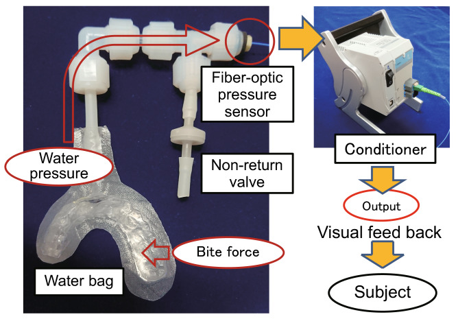
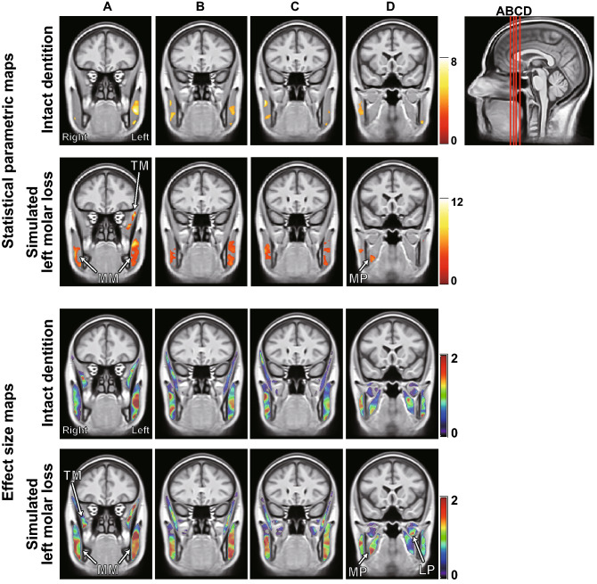
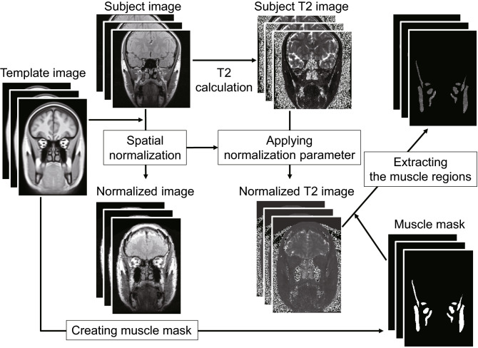
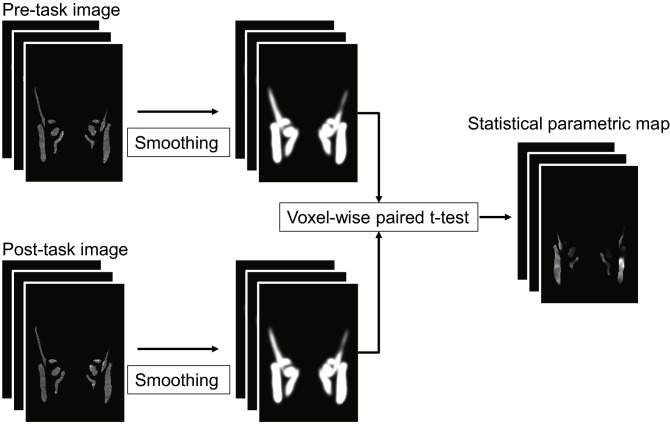
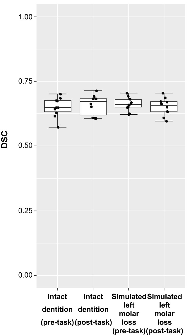
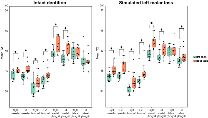
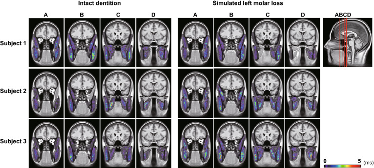
Analysis of the internal local activity distribution in human skeletal muscles is important for managing muscle fatigue/pain and dysfunction. However, no method is established for three-dimensional (3D) statistical analysis of features of activity regions common to multiple subjects during voluntary motor tasks. We investigated the characteristics of muscle activity distribution from the data of ten healthy subjects (29 ± 1 year old, 2 women) during voluntary teeth clenching under two different occlusal conditions by applying spatial normalization and statistical parametric mapping (SPM) to analysis of muscle functional magnetic resonance imaging (mfMRI) using increase in transverse relaxation time (T2) of the skeletal muscle induced by exercise. The expansion of areas with significant T2 increase was observed in the masticatory muscles after clenching with molar loss comparing with intact dentition. The muscle activity distribution characteristics common to a group of subjects, i.e., the active region in the temporal muscle ipsilateral to the side with the molar loss and medial pterygoid muscle contralateral to the side with the molar loss, were clarified in 3D by applying spatial normalization and SPM to mfMRI analysis. This method might elucidate the functional distribution within the muscles and the localized muscular activity related to skeletal muscle disorders.
分析人体骨骼肌的内部局部活动分布对于管理肌肉疲劳/疼痛和功能障碍很重要。然而,对于在自愿运动任务期间共同涉及多个受试者的活动区域的特征的三维(3D)统计分析,尚无方法建立。我们通过对肌肉功能磁共振成像(mfMRI)进行空间归一化和统计参数映射(SPM)分析,应用运动引起的骨骼肌横向弛豫时间(T2)增加,研究了十名健康受试者(29 ± 1 岁,2 名女性)在两种不同咬合条件下自愿咬牙时的肌肉活动分布特征。与完整的牙列相比,磨牙缺失后咬牙时咀嚼肌中 T2 增加的显著区域扩大。通过对 mfMRI 分析应用空间归一化和 SPM,我们在 3D 中阐明了一组受试者共同的肌肉活动分布特征,即在磨牙缺失侧的颞肌和对侧的翼内肌同侧的活动区域。这种方法可能阐明与骨骼肌疾病相关的肌肉内功能分布和局部肌肉活动。