Department of Pathology, Emory University School of Medicine, Atlanta, GA, USA.
Department of Pathology, University of Rochester Medical Center, Rochester, NY, USA.
Diagn Pathol. 2021 Feb 27;16(1):18. doi: 10.1186/s13000-021-01076-5.
Metastases are common in non-cirrhotic livers but are considered unlikely in the setting of cirrhosis. However, the degree of fibrosis in cirrhosis may vary; thus metastases may still access the liver vasculature and present as a mass in cirrhotic livers. This possibility may affect pathologists' diagnostic algorithms when faced with a liver mass biopsy.
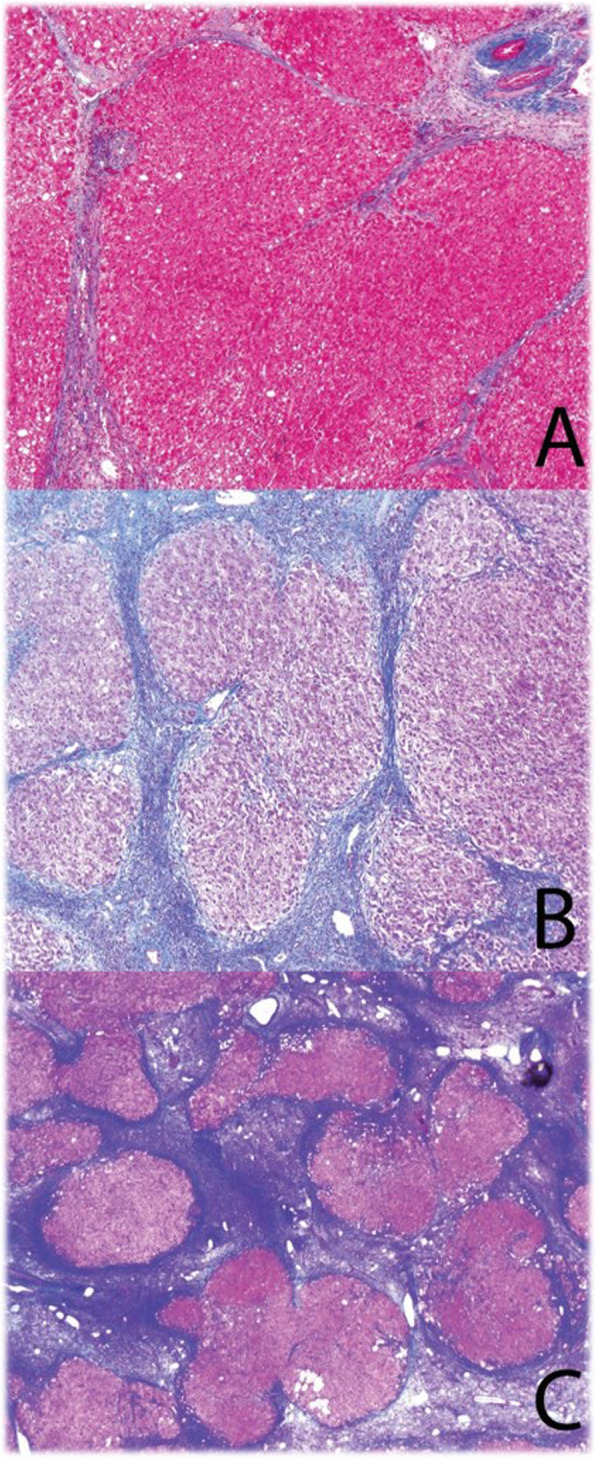
We hypothesized that metastases can occur in cirrhotic livers if fibrous remodeling is not severe or abnormal veno-arterial shunting exists to override an obstructed portal system. We searched departmental archives for cirrhotic livers with masses, categorizing fibrosis by Laennec staging: 4A = mild cirrhosis, 4B = moderate, 4 C = severe.
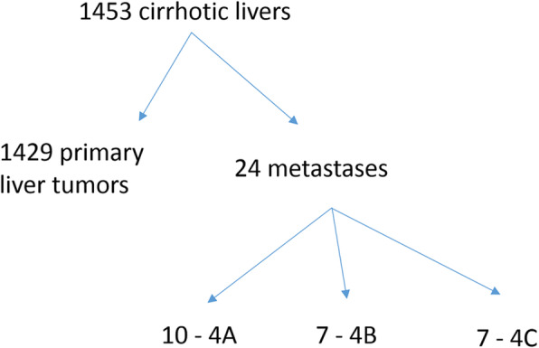
Of 1453 cirrhotic livers with masses, 1429 were primary tumors and 24 were metastases (1.7 %). Of livers with metastases, most had 4A or 4B cirrhosis by Laennec staging (n = 17; 71 %). Eleven patients were evaluated by ultrasound Doppler; 2 of 5 with Laennec 4 C had reversal of portal vein flow, but all 4A & 4B patients had patent portal veins without reversed flow. Echocardiograms (13 patients) showed no ventricular or atrial septal defects or arteriovenous shunts.
Metastases are uncommon in cirrhotic livers, accounting for 1.7 % of masses. Most involved livers had mild or moderate cirrhosis (Laennec 4A/4B) and patent portal veins; however, as some Laennec 4 C cases also contained metastases, obstructed portal access may not be enough to deter metastatic access.
转移瘤在非肝硬化肝脏中很常见,但在肝硬化中则不太可能发生。然而,肝硬化的纤维化程度可能会有所不同;因此,转移瘤仍可能进入肝血管系统,并在肝硬化肝脏中表现为肿块。这种可能性可能会影响病理学家在面对肝脏肿块活检时的诊断算法。
我们假设,如果纤维重塑不严重或存在异常的动静脉分流以克服阻塞的门脉系统,转移瘤仍可能发生在肝硬化肝脏中。我们在科室档案中搜索了有肿块的肝硬化肝脏,根据 Laennec 分期对纤维化进行分类:4A=轻度肝硬化,4B=中度,4C=重度。
在 1453 例有肿块的肝硬化肝脏中,1429 例为原发性肿瘤,24 例为转移瘤(1.7%)。在有转移瘤的肝脏中,大多数 Laennec 分期为 4A 或 4B 肝硬化(n=17;71%)。11 例患者接受了超声多普勒检查;5 例 Laennec 4C 中有 2 例门静脉血流逆转,但所有 4A 和 4B 患者的门静脉均通畅且无血流逆转。超声心动图(13 例)未显示室间隔或房间隔缺损或动静脉分流。
转移瘤在肝硬化肝脏中并不常见,占肿块的 1.7%。大多数受累肝脏的纤维化程度为轻度或中度(Laennec 4A/4B)且门静脉通畅;然而,由于一些 Laennec 4C 病例也包含转移瘤,因此阻塞的门脉通路可能不足以阻止转移瘤的进入。