• 文献检索
  • 文档翻译
  • 深度研究
  • 学术资讯
  • Suppr Zotero 插件Zotero 插件
  • 邀请有礼
  • 套餐&价格
  • 历史记录
应用&插件
Suppr Zotero 插件Zotero 插件浏览器插件Mac 客户端Windows 客户端微信小程序
定价
高级版会员购买积分包购买API积分包
服务
文献检索文档翻译深度研究API 文档MCP 服务
关于我们
关于 Suppr公司介绍联系我们用户协议隐私条款
关注我们

Suppr 超能文献

核心技术专利:CN118964589B侵权必究
粤ICP备2023148730 号-1Suppr @ 2026

文献检索

告别复杂PubMed语法,用中文像聊天一样搜索,搜遍4000万医学文献。AI智能推荐,让科研检索更轻松。

立即免费搜索

文件翻译

保留排版,准确专业,支持PDF/Word/PPT等文件格式,支持 12+语言互译。

免费翻译文档

深度研究

AI帮你快速写综述,25分钟生成高质量综述,智能提取关键信息,辅助科研写作。

立即免费体验

利用高分辨率多图像配准与平均技术(HR-MICRA)对海马内部结构进行清晰一致的成像。

Clear and Consistent Imaging of Hippocampal Internal Architecture With High Resolution Multiple Image Co-registration and Averaging (HR-MICRA).

作者信息

Ver Hoef Lawrence, Deshpande Hrishikesh, Cure Joel, Selladurai Goutham, Beattie Julia, Kennedy Richard E, Knowlton Robert C, Szaflarski Jerzy P

机构信息

Department of Neurology, The University of Alabama at Birmingham, Birmingham, AL, United States.

Department of Biomedical Engineering, The University of Alabama at Birmingham, Birmingham, AL, United States.

出版信息

Front Neurosci. 2021 Feb 11;15:546312. doi: 10.3389/fnins.2021.546312. eCollection 2021.

DOI:10.3389/fnins.2021.546312
PMID:33642971
原文链接:https://pmc.ncbi.nlm.nih.gov/articles/PMC7905096/
Abstract

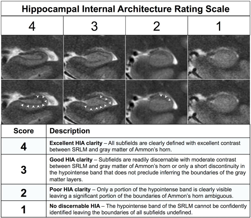
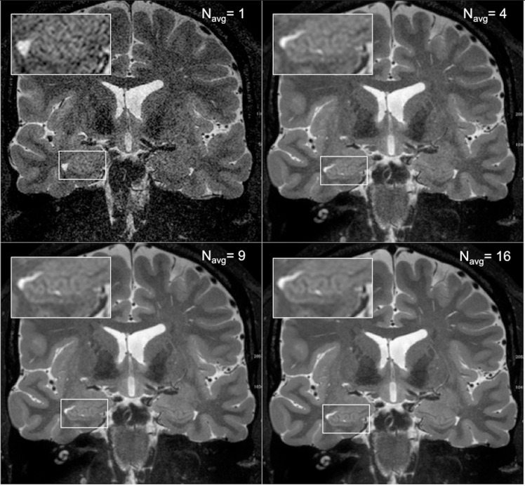
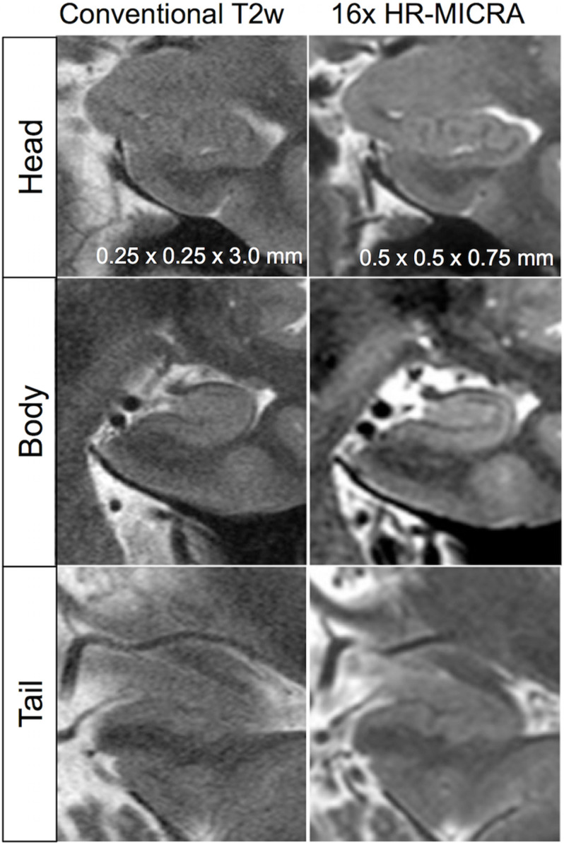
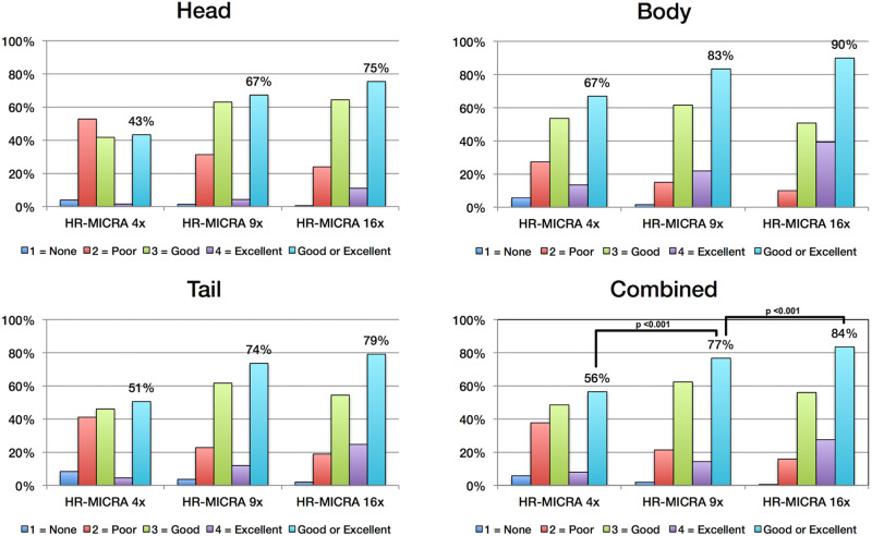
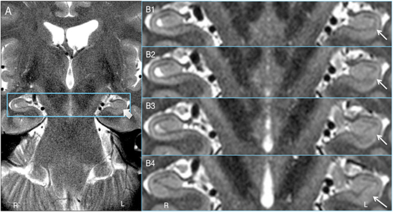
Magnetic resonance imaging of hippocampal internal architecture (HIA) at 3T is challenging. HIA is defined by layers of gray and white matter that are less than 1 mm thick in the coronal plane. To visualize HIA, conventional MRI approaches have relied on sequences with high in-plane resolution (≤0.5 mm) but comparatively thick slices (2-5 mm). However, thicker slices are prone to volume averaging effects that result in loss of HIA clarity and blurring of the borders of the hippocampal subfields in up to 61% of slices as has been reported. In this work we describe an approach to hippocampal imaging that provides consistently high HIA clarity using a commonly available sequence and post-processing techniques that is flexible and may be applicable to any MRI platform. We refer to this approach as High Resolution Multiple Image Co-registration and Averaging (HR-MICRA). This approach uses a variable flip angle turbo spin echo sequence to repeatedly acquire a whole brain T2w image volume with high resolution in three dimensions in a relatively short amount of time, and then co-register the volumes to correct for movement and average the repeated scans to improve SNR. We compared the averages of 4, 9, and 16 individual scans in 20 healthy controls using a published HIA clarity rating scale. In the body of the hippocampus, the proportion of slices with good or excellent HIA clarity was 90%, 83%, and 67% for the 16x, 9x, and 4x HR-MICRA images, respectively. Using the 4x HR-MICRA images as a baseline, the 9x HR-MICRA images were 2.6 times and 16x HR-MICRA images were 3.2 times more likely to have high HIA ratings ( < 0.001) across all hippocampal segments (head, body, and tail). The thin slices of the HR-MICRA images allow reformatting in any plane with clear visualization of hippocampal dentation in the sagittal plane. Clear and consistent visualization of HIA will allow application of this technique to future hippocampal structure research, as well as more precise manual or automated segmentation.

摘要

在3T条件下对海马体内部结构(HIA)进行磁共振成像具有挑战性。HIA由冠状面中厚度小于1毫米的灰质和白质层所界定。为了可视化HIA,传统的MRI方法依赖于具有高平面分辨率(≤0.5毫米)但切片相对较厚(2 - 5毫米)的序列。然而,较厚的切片容易出现容积平均效应,导致HIA清晰度下降,海马亚区边界模糊,据报道高达61%的切片会出现这种情况。在这项工作中,我们描述了一种海马成像方法,该方法使用常用序列和后处理技术,能始终如一地提供高清晰度的HIA,具有灵活性,且可能适用于任何MRI平台。我们将这种方法称为高分辨率多图像配准与平均(HR - MICRA)。这种方法使用可变翻转角快速自旋回波序列,在相对较短的时间内三维重复采集具有高分辨率的全脑T2加权图像容积,然后对这些容积进行配准以校正运动,并对重复扫描进行平均以提高信噪比。我们使用已发表的HIA清晰度评级量表,比较了20名健康对照者4次、9次和16次个体扫描的平均值。在海马体主体部分,1×、9×和4×HR - MICRA图像中HIA清晰度良好或优秀的切片比例分别为90%、83%和67%。以4×HR - MICRA图像作为基线,在所有海马段(头部、主体和尾部),9×HR - MICRA图像具有高HIA评级(<0.001)的可能性是4×HR - MICRA图像的2.6倍,16×HR - MICRA图像是其3.2倍。HR - MICRA图像的薄片允许在任何平面进行重新格式化,矢状面中海马齿状结构清晰可见。HIA清晰且一致的可视化将使该技术能够应用于未来的海马结构研究,以及更精确的手动或自动分割。

https://cdn.ncbi.nlm.nih.gov/pmc/blobs/a35a/7905096/1c39774d0d7c/fnins-15-546312-g008.jpg
https://cdn.ncbi.nlm.nih.gov/pmc/blobs/a35a/7905096/ca949ddfe92a/fnins-15-546312-g001.jpg
https://cdn.ncbi.nlm.nih.gov/pmc/blobs/a35a/7905096/8db8ede34adb/fnins-15-546312-g002.jpg
https://cdn.ncbi.nlm.nih.gov/pmc/blobs/a35a/7905096/ef7841cb7d7a/fnins-15-546312-g003.jpg
https://cdn.ncbi.nlm.nih.gov/pmc/blobs/a35a/7905096/486ce779eb43/fnins-15-546312-g004.jpg
https://cdn.ncbi.nlm.nih.gov/pmc/blobs/a35a/7905096/78cf3f9812d7/fnins-15-546312-g005.jpg
https://cdn.ncbi.nlm.nih.gov/pmc/blobs/a35a/7905096/01b5ca02fbdf/fnins-15-546312-g006.jpg
https://cdn.ncbi.nlm.nih.gov/pmc/blobs/a35a/7905096/f50f74a2133e/fnins-15-546312-g007.jpg
https://cdn.ncbi.nlm.nih.gov/pmc/blobs/a35a/7905096/1c39774d0d7c/fnins-15-546312-g008.jpg

相似文献

1
Clear and Consistent Imaging of Hippocampal Internal Architecture With High Resolution Multiple Image Co-registration and Averaging (HR-MICRA).利用高分辨率多图像配准与平均技术(HR-MICRA)对海马内部结构进行清晰一致的成像。
Front Neurosci. 2021 Feb 11;15:546312. doi: 10.3389/fnins.2021.546312. eCollection 2021.
2
Evaluating hippocampal internal architecture on MRI: inter-rater reliability of a proposed scoring system.评估 MRI 中海马内部结构:一个提出的评分系统的组内可靠性。
Epilepsy Res. 2013 Sep;106(1-2):146-54. doi: 10.1016/j.eplepsyres.2013.05.009. Epub 2013 Aug 3.
3
Multi-atlas segmentation of the whole hippocampus and subfields using multiple automatically generated templates.使用多个自动生成的模板对整个海马体及其子区进行多图谱分割。
Neuroimage. 2014 Nov 1;101:494-512. doi: 10.1016/j.neuroimage.2014.04.054. Epub 2014 Apr 29.
4
Study of the hippocampal internal architecture in temporal lobe epilepsy using 7 T and 3 T MRI.使用 7T 和 3T MRI 研究颞叶癫痫的海马内结构。
Seizure. 2019 Oct;71:116-123. doi: 10.1016/j.seizure.2019.06.023. Epub 2019 Jul 9.
5
Curved multiplanar reformatting provides improved visualization of hippocampal anatomy.曲面多平面重建可改善海马解剖结构的可视化效果。
Hippocampus. 2020 Feb;30(2):156-161. doi: 10.1002/hipo.23177. Epub 2019 Nov 19.
6
Volumetric comparison of hippocampal subfields extracted from 4-minute accelerated vs. 8-minute high-resolution T2-weighted 3T MRI scans.从 4 分钟加速与 8 分钟高分辨率 3T MRI 扫描中提取的海马亚区的容积比较。
Brain Imaging Behav. 2018 Dec;12(6):1583-1595. doi: 10.1007/s11682-017-9819-3.
7
A novel in vivo atlas of human hippocampal subfields using high-resolution 3 T magnetic resonance imaging.利用高分辨率 3T 磁共振成像构建人类海马亚区的新型活体图谱。
Neuroimage. 2013 Jul 1;74:254-65. doi: 10.1016/j.neuroimage.2013.02.003. Epub 2013 Feb 13.
8
Longitudinal reproducibility of automatically segmented hippocampal subfields: A multisite European 3T study on healthy elderly.自动分割海马亚区的纵向可重复性:一项针对健康老年人的欧洲多中心3T研究。
Hum Brain Mapp. 2015 Sep;36(9):3516-27. doi: 10.1002/hbm.22859. Epub 2015 Jun 3.
9
In vivo quantification of hippocampal subfields using 4.7 T fast spin echo imaging.使用 4.7T 快速自旋回波成像进行海马亚区的体内定量分析。
Neuroimage. 2010 Jan 15;49(2):1224-30. doi: 10.1016/j.neuroimage.2009.09.042. Epub 2009 Sep 26.
10
Edge-enhancing gradient echo with multi-image co-registration and averaging (EDGE-MICRA) for targeting thalamic centromedian and parafascicular nuclei.用于靶向丘脑中央中核和丘脑束旁核的边缘增强梯度回波多图像配准和平均(EDGE-MICRA)。
Neuroradiol J. 2021 Dec;34(6):667-675. doi: 10.1177/19714009211021781. Epub 2021 Jun 13.

引用本文的文献

1
Segmentation of hippocampal subregion via 5-Tesla magnetic resonance imaging: a comparative study.通过5特斯拉磁共振成像对海马亚区进行分割:一项比较研究。
Quant Imaging Med Surg. 2025 May 1;15(5):3861-3874. doi: 10.21037/qims-24-2169. Epub 2025 Apr 10.
2
Effects of a brief HIIT intervention on cognitive performance in older women.短时间高强度间歇训练对老年女性认知表现的影响。
Geroscience. 2024 Feb;46(1):1371-1384. doi: 10.1007/s11357-023-00893-4. Epub 2023 Aug 15.
3
Development of an objective method to quantify hippocampal dentation.

本文引用的文献

1
Non-linear realignment improves hippocampus subfield segmentation reliability.非线性重配可提高海马亚区分割的可靠性。
Neuroimage. 2019 Dec;203:116206. doi: 10.1016/j.neuroimage.2019.116206. Epub 2019 Sep 17.
2
Accuracy of the Compressed Sensing Accelerated 3D-FLAIR Sequence for the Detection of MS Plaques at 3T.压缩感知加速 3D-FLAIR 序列在 3T 下检测 MS 斑块的准确性。
AJNR Am J Neuroradiol. 2018 Mar;39(3):454-458. doi: 10.3174/ajnr.A5517. Epub 2018 Jan 18.
3
Development of a histologically validated segmentation protocol for the hippocampal body.
开发一种客观的方法来量化海马齿状回。
Hum Brain Mapp. 2023 Jun 1;44(8):2967-2980. doi: 10.1002/hbm.26222. Epub 2023 Mar 27.
4
Recent Advances in Neuroimaging of Epilepsy.癫痫的神经影像学最新进展。
Neurotherapeutics. 2021 Apr;18(2):811-826. doi: 10.1007/s13311-021-01049-y. Epub 2021 May 3.
开发一种基于组织学验证的海马体分割协议。
Neuroimage. 2017 Aug 15;157:219-232. doi: 10.1016/j.neuroimage.2017.06.008. Epub 2017 Jun 3.
4
Hippocampal dentation: Structural variation and its association with episodic memory in healthy adults.海马齿状回:健康成年人的结构变异及其与情景记忆的关联。
Neuropsychologia. 2017 Jul 1;101:65-75. doi: 10.1016/j.neuropsychologia.2017.04.036. Epub 2017 May 1.
5
Aberrant Hippocampal Connectivity in Unmedicated Patients With Schizophrenia and Effects of Antipsychotic Medication: A Longitudinal Resting State Functional MRI Study.未用药精神分裂症患者的海马体连接异常及抗精神病药物的影响:一项纵向静息态功能磁共振成像研究
Schizophr Bull. 2016 Jul;42(4):1046-55. doi: 10.1093/schbul/sbv228. Epub 2016 Feb 12.
6
Automated Hippocampal Subfield Segmentation at 7T MRI.7T磁共振成像下的海马亚区自动分割
AJNR Am J Neuroradiol. 2016 Jun;37(6):1050-7. doi: 10.3174/ajnr.A4659. Epub 2016 Feb 4.
7
Simultaneous multislice (SMS) imaging techniques.同时多层(SMS)成像技术。
Magn Reson Med. 2016 Jan;75(1):63-81. doi: 10.1002/mrm.25897. Epub 2015 Aug 26.
8
A computational atlas of the hippocampal formation using ex vivo, ultra-high resolution MRI: Application to adaptive segmentation of in vivo MRI.利用离体超高分辨率磁共振成像构建的海马结构计算图谱:在活体磁共振成像自适应分割中的应用
Neuroimage. 2015 Jul 15;115:117-37. doi: 10.1016/j.neuroimage.2015.04.042. Epub 2015 Apr 29.
9
Quantitative comparison of 21 protocols for labeling hippocampal subfields and parahippocampal subregions in in vivo MRI: towards a harmonized segmentation protocol.21种用于在活体磁共振成像中标记海马亚区和海马旁回亚区域的方案的定量比较:迈向统一的分割方案
Neuroimage. 2015 May 1;111:526-41. doi: 10.1016/j.neuroimage.2015.01.004. Epub 2015 Jan 14.
10
A Critical Appraisal of the Hippocampal Subfield Segmentation Package in FreeSurfer.对FreeSurfer中海马亚区分割软件包的批判性评估。
Front Aging Neurosci. 2014 Sep 25;6:261. doi: 10.3389/fnagi.2014.00261. eCollection 2014.