Division of Neurology, Department of Medicine, University of Alberta, Edmonton, Alberta, Canada.
Department of Biomedical Engineering, University of Alberta, Edmonton, Alberta, Canada.
Hippocampus. 2020 Feb;30(2):156-161. doi: 10.1002/hipo.23177. Epub 2019 Nov 19.
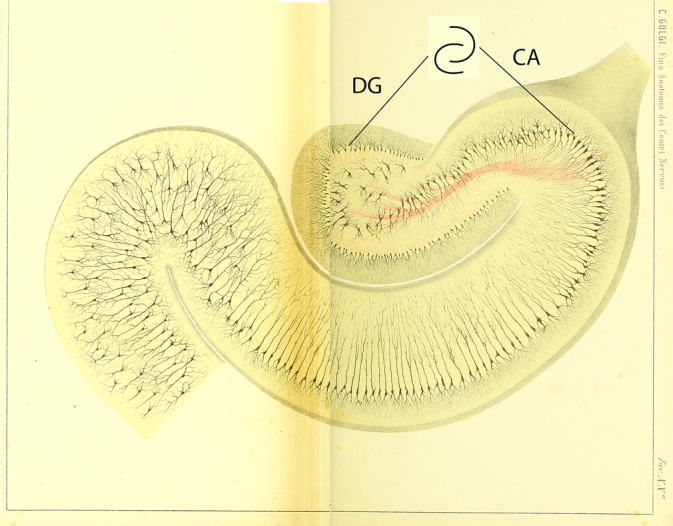
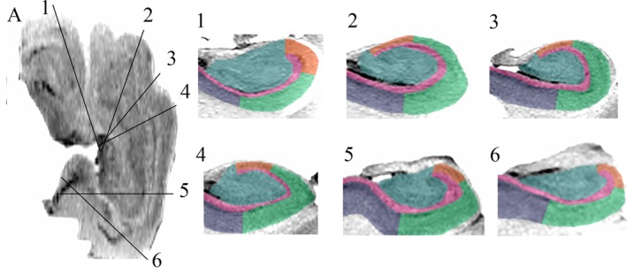
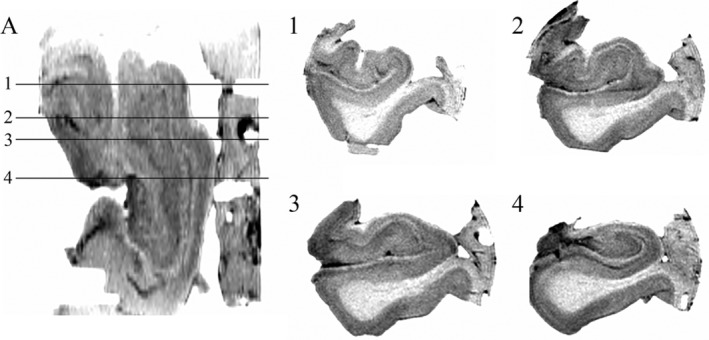
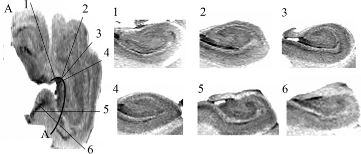
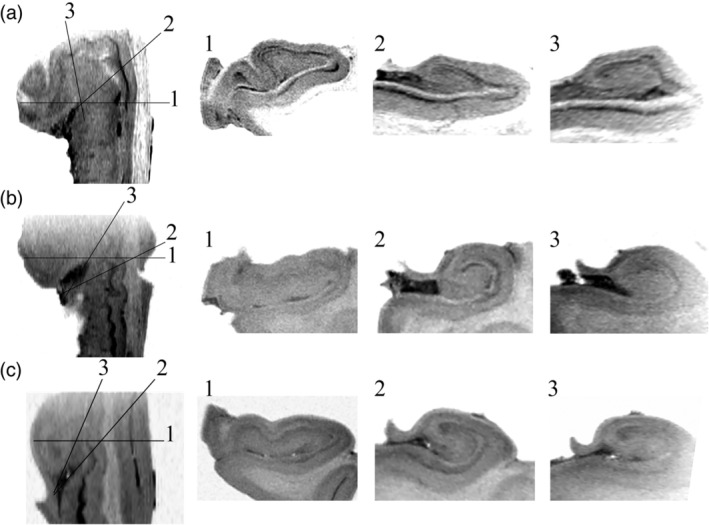
There is a growing body of literature studying changes in hippocampal subfields in a variety of different neurological conditions, but this work has mainly focused on the hippocampal body given challenges in visualization of hippocampal anatomy in the head and tail when sectioned in the typical coronal image plane. Curved multiplanar reformatting (CMPR) is an image reconstruction method that can improve visualization of complex three-dimensional structures. The objective of this study was to determine whether CMPR could facilitate visualization of the human hippocampal anatomy along the entire caudal-rostral axis. CMPR was applied to high-resolution magnetic resonance imaging acquired ex vivo on four cadaveric hippocampal specimens at 4.7 T (T2-weighted, 0.2 × 0.2 × 0.5 mm ). CMPR provided clear visualization of the classic "interlocking C" appearance of the dentate gyrus and cornu ammonis along the entire caudal-rostral axis including the head and tail, which otherwise show complex anatomy on the standard coronal slices. CMPR facilitated visualization of hippocampal anatomy providing the impetus to develop simplified approaches to delineate subfields along the entire hippocampus including the usually neglected head and tail.
越来越多的文献研究了各种不同神经状况下海马亚区的变化,但这项工作主要集中在海马体上,因为在典型的冠状影像平面上进行切片时,头部和尾部的海马解剖结构难以可视化。曲面多平面重建成像(CMPR)是一种可以改善复杂三维结构可视化的图像重建方法。本研究的目的是确定 CMPR 是否可以促进沿着整个头尾轴可视化人类海马解剖结构。CMPR 应用于在 4.7T 下离体采集的四个尸体海马标本的高分辨率磁共振成像(T2 加权,0.2×0.2×0.5mm)。CMPR 清晰地显示了齿状回和角回的经典“互锁 C”外观,沿着整个头尾轴,包括头部和尾部,而在标准冠状切片上,这些部位显示出复杂的解剖结构。CMPR 促进了海马解剖结构的可视化,为开发简化的方法来描绘整个海马体(包括通常被忽视的头部和尾部)的亚区提供了动力。