Bruder Jonas C, Schmelzeisen Christoph, Lachner-Piza Daniel, Reinacher Peter, Schulze-Bonhage Andreas, Jacobs Julia
Department of Neuropediatrics and Muscular Disease, University Medical Center, Freiburg, Germany.
Stereotactic & Functional Neurosurgery, University Medical Center, Freiburg, Germany.
Front Neurol. 2021 Feb 10;12:612293. doi: 10.3389/fneur.2021.612293. eCollection 2021.
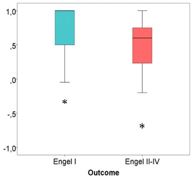
High frequency oscillations (HFO) are promising biomarkers of epileptic tissue. While group analysis suggested a correlation between surgical removal of HFO generating tissue and seizure free outcome, HFO could not predict seizure outcome on an individual patient level. One possible explanation is the lack of differentiation between physiological and epileptic HFO. In the mesio-temporal lobe, a proportion of physiological ripples can be identified by their association with scalp sleep spindles. Spike associated ripples in contrast can be considered epileptic. This study investigated whether categorizing ripples by the co-occurrence with sleep spindles or spikes improves outcome prediction after surgery. Additionally, it aimed to investigate whether spindle-ripple association is limited to the mesio-temporal lobe structures or visible across the whole brain. We retrospectively analyzed EEG of 31 patients with chronic intracranial EEG. Sleep spindles in scalp EEG and ripples and epileptic spikes in iEEG were automatically detected. Three ripple subtypes were obtained: SpindleR, Non-SpindleR, and SpikeR. Rate ratios between removed and non-removed brain areas were calculated. We compared the distinct ripple subtypes and their rates in different brain regions, inside and outside seizure onset areas and between patients with good and poor seizure outcome. SpindleR were found across all brain regions. SpikeR had significantly higher rates in the SOZ than in Non-SOZ channels. A significant positive correlation between removal of ripple-events and good outcome was found for the mixed ripple group (r = 0.43, = 0.017) and for ripples not associated with spindles (r=0.40, = 0.044). Also, a significantly high proportion of spikes associated with ripples were removed in seizure free patients ( = 0.036). SpindleR are found in mesio-temporal and neocortical structures, indicating that ripple-spindle-coupling might have functional importance beyond mesio-temporal structures. Overall, the proportion of SpindleR was low and separating spindle and spike associated ripples did not improve outcome prediction in our patient group. SpindleR analysis therefore can be a tool to identify physiological events but needs to be used in combination with other methods to have clinical relevance.
高频振荡(HFO)是癫痫组织很有前景的生物标志物。虽然群体分析表明切除产生HFO的组织与无癫痫发作结果之间存在相关性,但HFO无法在个体患者层面预测癫痫发作结果。一种可能的解释是生理HFO和癫痫性HFO之间缺乏区分。在颞叶内侧,一部分生理涟漪可通过其与头皮睡眠纺锤波的关联来识别。相比之下,与棘波相关的涟漪可被视为癫痫性的。本研究调查了根据与睡眠纺锤波或棘波的同时出现对涟漪进行分类是否能改善手术后的结果预测。此外,其旨在研究纺锤波 - 涟漪关联是否仅限于颞叶内侧结构,还是在全脑可见。我们回顾性分析了31例慢性颅内脑电图患者的脑电图。自动检测头皮脑电图中的睡眠纺锤波以及颅内脑电图中的涟漪和癫痫棘波。获得了三种涟漪亚型:纺锤波相关涟漪(SpindleR)、非纺锤波相关涟漪(Non-SpindleR)和棘波相关涟漪(SpikeR)。计算切除和未切除脑区之间的率比。我们比较了不同脑区、癫痫发作起始区域内外以及癫痫发作结果良好和不良患者之间不同的涟漪亚型及其发生率。在所有脑区均发现了纺锤波相关涟漪。棘波相关涟漪在癫痫发作起始区(SOZ)的发生率显著高于非癫痫发作起始区通道。对于混合涟漪组(r = 0.43,P = 0.017)以及与纺锤波无关的涟漪(r = 0.40,P = 0.044),发现切除涟漪事件与良好结果之间存在显著正相关。此外,在无癫痫发作患者中,与涟漪相关的棘波的切除比例也显著较高(P = 0.036)。在颞叶内侧和新皮质结构中均发现了纺锤波相关涟漪,这表明涟漪 - 纺锤波耦合可能在颞叶内侧结构之外具有功能重要性。总体而言,纺锤波相关涟漪的比例较低,在我们的患者群体中,区分与纺锤波和棘波相关的涟漪并不能改善结果预测。因此,纺锤波相关涟漪分析可以作为识别生理事件的一种工具,但需要与其他方法结合使用才有临床相关性。