• 文献检索
  • 文档翻译
  • 深度研究
  • 学术资讯
  • Suppr Zotero 插件Zotero 插件
  • 邀请有礼
  • 套餐&价格
  • 历史记录
应用&插件
Suppr Zotero 插件Zotero 插件浏览器插件Mac 客户端Windows 客户端微信小程序
定价
高级版会员购买积分包购买API积分包
服务
文献检索文档翻译深度研究API 文档MCP 服务
关于我们
关于 Suppr公司介绍联系我们用户协议隐私条款
关注我们

Suppr 超能文献

核心技术专利:CN118964589B侵权必究
粤ICP备2023148730 号-1Suppr @ 2026

文献检索

告别复杂PubMed语法,用中文像聊天一样搜索,搜遍4000万医学文献。AI智能推荐,让科研检索更轻松。

立即免费搜索

文件翻译

保留排版,准确专业,支持PDF/Word/PPT等文件格式,支持 12+语言互译。

免费翻译文档

深度研究

AI帮你快速写综述,25分钟生成高质量综述,智能提取关键信息,辅助科研写作。

立即免费体验

泪液血管生成因子:早产儿视网膜病变筛查的潜在非侵入性生物标志物。

Tear Fluid Angiogenic Factors: Potential Noninvasive Biomarkers for Retinopathy of Prematurity Screening in Preterm Infants.

机构信息

Department of Pediatric Retina, Narayana Nethralaya, Bangalore, India.

GROW Research Lab, Narayana Nethralaya Foundation, Bangalore, India.

出版信息

Invest Ophthalmol Vis Sci. 2021 Mar 1;62(3):2. doi: 10.1167/iovs.62.3.2.

DOI:10.1167/iovs.62.3.2
PMID:33646290
原文链接:https://pmc.ncbi.nlm.nih.gov/articles/PMC7938022/
Abstract

PURPOSE

To determine the status of proangiogenic factors in the tear fluid of preterm infants with and without retinopathy of prematurity (ROP).

METHODS

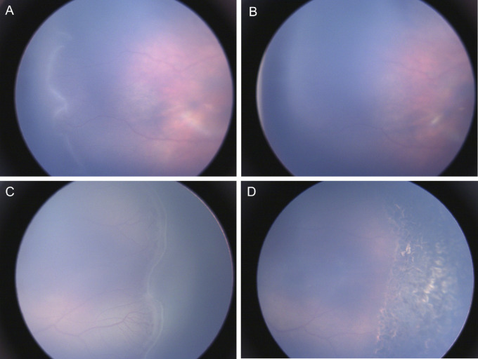
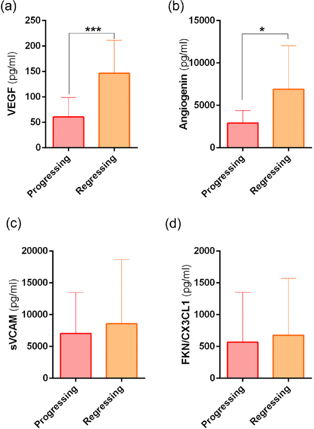
Preterm infants (n = 36) undergoing routine ROP screening included in the prospective study were categorized as No-ROP (n = 13, no ROP at any visits), ROP (if ROP was present at first visit; n = 18), or No-ROP to ROP (no disease at first visit, but developed ROP subsequently; n = 5). Infants with ROP were also grouped as progressing (n = 7) and regressing (n = 16) based on ROP evolution between the first and subsequent visits. Schirmer's strips were used to collect tear fluid and proangiogenic factors (VEGF, angiogenin, soluble vascular cell adhesion molecule, and fractalkine) levels (in picograms per milliliter) in tear fluid were measured by multiplex ELISA.

RESULTS

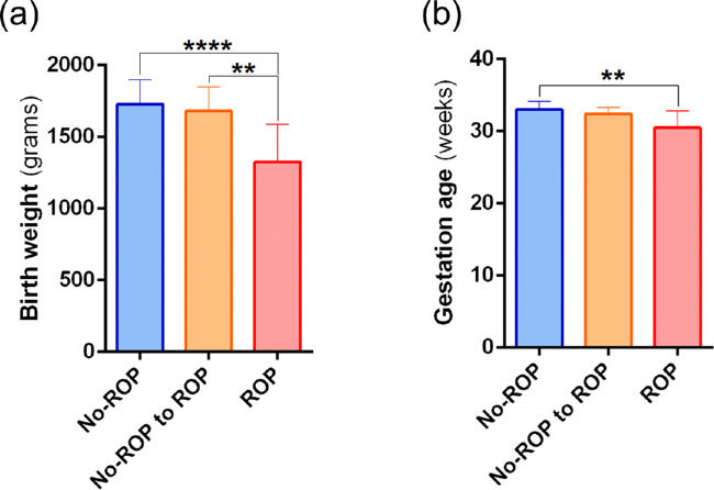
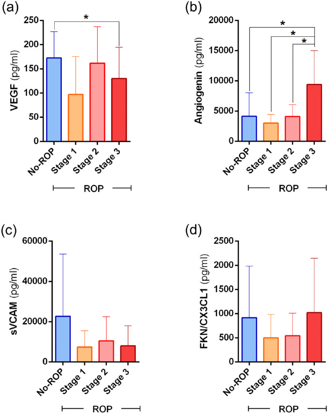
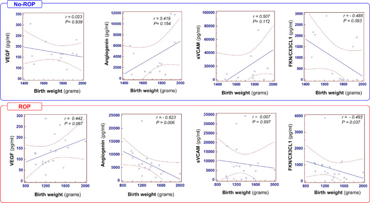
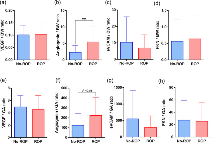
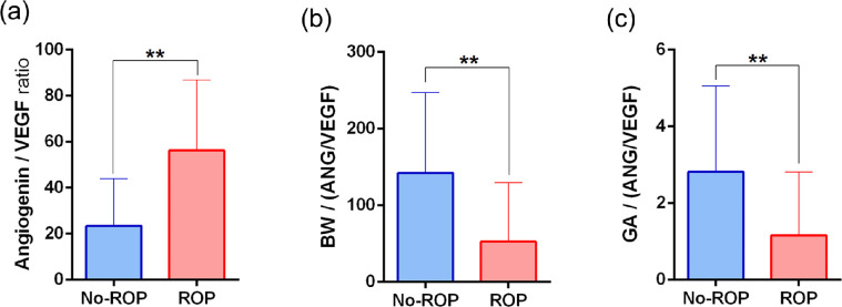
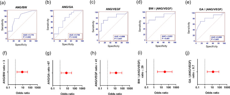
Lower levels of VEGF (135 ± 69; mean ± standard deviation) and higher levels of angiogenin (6568 ± 4975) were observed in infants with ROP compared with infants without ROP (172.5 ± 54.0; 4139 ± 3909) at the first visit. Significantly lower levels of VEGF were observed in the No-ROP to ROP group compared with the No-ROP and ROP groups. The VEGF and angiogenin levels at the first visit were significantly lower in infants with ROP with progressing disease. Angiogenin levels negatively correlated with birth weight and gestational age in ROP. The area under the curve (AUC) and odds ratio (OR) analysis demonstrated that angiogenin/birth weight (AUC = 0.776; OR, 8.6); angiogenin/gestational age (AUC = 0.706; OR, 7.3) and Angiogenin/VEGF (AUC = 0.806; OR, 14.3) ratios were able to differentiated preterm infants with and without ROP.

CONCLUSIONS

The association between angiogenin and ROP suggests its possible role in ROP. The ratio of angiogenin level with birth weight, gestational age, and/or VEGF could serve as a potential noninvasive screening biomarker for ROP.

摘要

目的

确定早产儿有无早产儿视网膜病变(ROP)的泪液中促血管生成因子的状态。

方法

纳入前瞻性研究的接受常规 ROP 筛查的早产儿(n=36)根据有无 ROP 分为无 ROP 组(n=13,各次就诊均无 ROP)、ROP 组(首次就诊时即存在 ROP,n=18)或无 ROP 至 ROP 组(首次就诊时无疾病,但随后发展为 ROP,n=5)。ROP 患儿还根据首次和随后就诊之间的 ROP 进展情况分为进展组(n=7)和消退组(n=16)。用 Schirmer 条收集泪液,用多重 ELISA 法测定泪液中促血管生成因子(VEGF、血管生成素、可溶性血管细胞黏附分子和 fractalkine)水平(以皮克/毫升为单位)。

结果

与无 ROP 组相比,首次就诊时 ROP 患儿的 VEGF 水平(135±69)较低,而血管生成素水平(6568±4975)较高(172.5±54.0,4139±3909)。无 ROP 至 ROP 组的 VEGF 水平显著低于无 ROP 组和 ROP 组。进展性 ROP 患儿首次就诊时 VEGF 和血管生成素水平显著较低。ROP 患儿的血管生成素水平与出生体重和胎龄呈负相关。ROC 曲线(AUC)和比值比(OR)分析表明,血管生成素/出生体重(AUC=0.776;OR,8.6)、血管生成素/胎龄(AUC=0.706;OR,7.3)和血管生成素/VEGF(AUC=0.806;OR,14.3)比值可区分有无 ROP 的早产儿。

结论

血管生成素与 ROP 之间的关联表明其可能在 ROP 中起作用。血管生成素水平与出生体重、胎龄和/或 VEGF 的比值可能成为 ROP 的潜在非侵入性筛查生物标志物。

https://cdn.ncbi.nlm.nih.gov/pmc/blobs/0f31/7938022/83d11df47b5b/iovs-62-3-2-f012.jpg
https://cdn.ncbi.nlm.nih.gov/pmc/blobs/0f31/7938022/98ed4b01a5d1/iovs-62-3-2-f001.jpg
https://cdn.ncbi.nlm.nih.gov/pmc/blobs/0f31/7938022/5f5da3041521/iovs-62-3-2-f002.jpg
https://cdn.ncbi.nlm.nih.gov/pmc/blobs/0f31/7938022/21ce3842a34f/iovs-62-3-2-f003.jpg
https://cdn.ncbi.nlm.nih.gov/pmc/blobs/0f31/7938022/a6924b6adef7/iovs-62-3-2-f004.jpg
https://cdn.ncbi.nlm.nih.gov/pmc/blobs/0f31/7938022/fb4e51be5d62/iovs-62-3-2-f005.jpg
https://cdn.ncbi.nlm.nih.gov/pmc/blobs/0f31/7938022/e8e82342c2f5/iovs-62-3-2-f006.jpg
https://cdn.ncbi.nlm.nih.gov/pmc/blobs/0f31/7938022/0ba849e8b381/iovs-62-3-2-f007.jpg
https://cdn.ncbi.nlm.nih.gov/pmc/blobs/0f31/7938022/34c8ef783e3c/iovs-62-3-2-f008.jpg
https://cdn.ncbi.nlm.nih.gov/pmc/blobs/0f31/7938022/641ab74c0197/iovs-62-3-2-f009.jpg
https://cdn.ncbi.nlm.nih.gov/pmc/blobs/0f31/7938022/0881020d1659/iovs-62-3-2-f010.jpg
https://cdn.ncbi.nlm.nih.gov/pmc/blobs/0f31/7938022/1cded50b1d47/iovs-62-3-2-f011.jpg
https://cdn.ncbi.nlm.nih.gov/pmc/blobs/0f31/7938022/83d11df47b5b/iovs-62-3-2-f012.jpg

相似文献

1
Tear Fluid Angiogenic Factors: Potential Noninvasive Biomarkers for Retinopathy of Prematurity Screening in Preterm Infants.泪液血管生成因子:早产儿视网膜病变筛查的潜在非侵入性生物标志物。
Invest Ophthalmol Vis Sci. 2021 Mar 1;62(3):2. doi: 10.1167/iovs.62.3.2.
2
Early detection and correlation of tear fluid inflammatory factors that influence angiogenesis in premature infants with and without retinopathy of prematurity.早期检测和相关性分析对早产儿有无早产儿视网膜病变影响血管生成的泪液炎症因子。
Indian J Ophthalmol. 2023 Nov;71(11):3465-3472. doi: 10.4103/IJO.IJO_3407_22.
3
Tear fluid cytokine analysis: a non-invasive approach for assessing retinopathy of prematurity severity.泪液细胞因子分析:评估早产儿视网膜病变严重程度的非侵入性方法。
Jpn J Ophthalmol. 2024 Sep;68(5):482-489. doi: 10.1007/s10384-024-01084-0. Epub 2024 Jul 10.
4
Tear Proteomics in Infants at Risk of Retinopathy of Prematurity: A Feasibility Study.早产儿视网膜病变风险婴儿的泪液蛋白质组学:一项可行性研究。
Transl Vis Sci Technol. 2024 May 1;13(5):1. doi: 10.1167/tvst.13.5.1.
5
Inflammatory and Angiogenic Mediators in Amniotic Fluid Are Associated With the Development of Retinopathy of Prematurity in Preterm Infants.羊水中的炎症和血管生成介质与早产儿视网膜病变的发展有关。
Invest Ophthalmol Vis Sci. 2020 May 11;61(5):42. doi: 10.1167/iovs.61.5.42.
6
Cord blood transforming growth factor-β-induced as predictive biomarker of retinopathy of prematurity in preterm infants.脐带血转化生长因子-β诱导作为早产儿视网膜病变的预测生物标志物。
Graefes Arch Clin Exp Ophthalmol. 2023 Sep;261(9):2477-2488. doi: 10.1007/s00417-023-06056-7. Epub 2023 Apr 6.
7
Quantitative analysis of tear angiogenic factors in retinopathy of prematurity: a pilot biomarker study.早产儿视网膜病变中泪液血管生成因子的定量分析:一项生物标志物初步研究
J AAPOS. 2023 Feb;27(1):14.e1-14.e6. doi: 10.1016/j.jaapos.2022.10.007. Epub 2022 Dec 26.
8
Correlation between tear levels of vascular endothelial growth factor and vitamin D at retinopathy of prematurity stages in preterm infants.早产儿视网膜病变各期泪液血管内皮生长因子与维生素 D 的相关性研究。
Sci Rep. 2023 Sep 27;13(1):16175. doi: 10.1038/s41598-023-43338-w.
9
Immune and Inflammatory Proteins in Cord Blood as Predictive Biomarkers of Retinopathy of Prematurity in Preterm Infants.脐血中免疫和炎症蛋白作为早产儿视网膜病变的预测生物标志物。
Invest Ophthalmol Vis Sci. 2019 Sep 3;60(12):3813-3820. doi: 10.1167/iovs.19-27258.
10
VEGF-A, VEGFR-1, VEGFR-2 and Tie2 levels in plasma of premature infants: relationship to retinopathy of prematurity.早产儿血浆中血管内皮生长因子A、血管内皮生长因子受体-1、血管内皮生长因子受体-2和Tie2水平:与早产儿视网膜病变的关系
Br J Ophthalmol. 2008 May;92(5):689-93. doi: 10.1136/bjo.2007.128371. Epub 2008 Apr 11.

引用本文的文献

1
Retinopathy of Prematurity (ROP): An Overview of Biomarkers in Various Samples for Prediction, Diagnosis, and Prognosis.早产儿视网膜病变(ROP):用于预测、诊断和预后的各种样本中生物标志物概述
Clin Ophthalmol. 2025 May 7;19:1515-1530. doi: 10.2147/OPTH.S519292. eCollection 2025.
2
Identifying potential tear biomarkers in premature infants with retinopathy of prematurity based on proteome and transcriptome analysis.基于蛋白质组和转录组分析鉴定早产儿视网膜病变潜在的泪液生物标志物。
Graefes Arch Clin Exp Ophthalmol. 2025 May 9. doi: 10.1007/s00417-025-06838-1.
3
Analysis of Salivary Cytokines in Retinopathy of Prematurity.

本文引用的文献

1
Intravitreal antivascular endothelial growth factor injection versus laser photocoagulation for retinopathy of prematurity: A meta-analysis of 3,701 eyes.玻璃体腔内抗血管内皮生长因子注射与激光光凝治疗早产儿视网膜病变:3701 只眼的荟萃分析。
Surv Ophthalmol. 2021 Jul-Aug;66(4):572-584. doi: 10.1016/j.survophthal.2020.12.002. Epub 2020 Dec 16.
2
A proposal of an algorithm for the diagnosis and treatment of recurrence or treatment failure of retinopathy of prematurity after anti-VEGF therapy based on a large case series.一项基于大病例系列的抗 VEGF 治疗后早产儿视网膜病变复发或治疗失败的诊断和治疗算法的建议。
Graefes Arch Clin Exp Ophthalmol. 2020 Apr;258(4):767-772. doi: 10.1007/s00417-020-04605-y. Epub 2020 Jan 17.
3
早产儿视网膜病变唾液细胞因子分析
Children (Basel). 2025 Jan 10;12(1):80. doi: 10.3390/children12010080.
4
The association of cytokine levels and postnatal factors with retinopathy of prematurity.细胞因子水平及产后因素与早产儿视网膜病变的关联
Indian J Ophthalmol. 2025 Jan 1;73(Suppl 1):S139-S143. doi: 10.4103/IJO.IJO_515_24. Epub 2024 Oct 25.
5
Comprehensive analysis of systemic, metabolic, and molecular changes following prospective change to low-carbohydrate diet in adults with type 2 diabetes mellitus in India.对印度2型糖尿病成年患者前瞻性采用低碳水化合物饮食后全身、代谢及分子变化的综合分析。
Front Nutr. 2024 Aug 30;11:1394298. doi: 10.3389/fnut.2024.1394298. eCollection 2024.
6
Circulating VEGF-A Levels in Relation to Retinopathy of Prematurity and Treatment Effects: A Systematic Review and Meta-Analysis.循环血管内皮生长因子A水平与早产儿视网膜病变及治疗效果的关系:一项系统评价和荟萃分析
Ophthalmol Sci. 2024 May 7;4(6):100548. doi: 10.1016/j.xops.2024.100548. eCollection 2024 Nov-Dec.
7
Tear fluid cytokine analysis: a non-invasive approach for assessing retinopathy of prematurity severity.泪液细胞因子分析:评估早产儿视网膜病变严重程度的非侵入性方法。
Jpn J Ophthalmol. 2024 Sep;68(5):482-489. doi: 10.1007/s10384-024-01084-0. Epub 2024 Jul 10.
8
Retinal Blood Vessel Formation in the Macula Following Intravitreal Ranibizumab Injection for Aggressive Retinopathy of Prematurity.玻璃体内注射雷珠单抗治疗侵袭性早产儿视网膜病变后黄斑区视网膜血管形成
Cureus. 2024 May 9;16(5):e60005. doi: 10.7759/cureus.60005. eCollection 2024 May.
9
Optical coherence tomography biomarkers in preterm infants with and without retinopathy of prematurity: fallacies and facts.患有和未患早产儿视网膜病变的早产儿的光学相干断层扫描生物标志物:谬误与事实
Eye (Lond). 2024 Sep;38(13):2656-2657. doi: 10.1038/s41433-024-03061-w. Epub 2024 Apr 30.
10
Tear Proteomics in Infants at Risk of Retinopathy of Prematurity: A Feasibility Study.早产儿视网膜病变风险婴儿的泪液蛋白质组学:一项可行性研究。
Transl Vis Sci Technol. 2024 May 1;13(5):1. doi: 10.1167/tvst.13.5.1.
Retinopathy of prematurity: incidence report of outliers based on international screening guidelines.
早产儿视网膜病变:基于国际筛查指南的异常值发病率报告
Int J Retina Vitreous. 2019 Dec 12;5(Suppl 1):53. doi: 10.1186/s40942-019-0203-x. eCollection 2019.
4
Comprehensive analysis of vitreous chemokines involved in ischemic retinal vein occlusion.缺血性视网膜静脉阻塞中玻璃体内趋化因子的综合分析
Mol Vis. 2019 Nov 15;25:756-765. eCollection 2019.
5
A review of treatment for retinopathy of prematurity.早产儿视网膜病变的治疗综述。
Expert Rev Ophthalmol. 2019;14(2):73-87. doi: 10.1080/17469899.2019.1596026. Epub 2019 Mar 29.
6
Retinopathy of prematurity treatment: Asian perspectives.早产儿视网膜病变治疗:亚洲视角。
Eye (Lond). 2020 Apr;34(4):632-642. doi: 10.1038/s41433-019-0643-4. Epub 2019 Oct 29.
7
Ranibizumab versus laser therapy for the treatment of very low birthweight infants with retinopathy of prematurity (RAINBOW): an open-label randomised controlled trial.雷珠单抗与激光治疗用于治疗极低出生体重早产儿视网膜病变(RAINBOW):一项开放标签随机对照试验。
Lancet. 2019 Oct 26;394(10208):1551-1559. doi: 10.1016/S0140-6736(19)31344-3. Epub 2019 Sep 12.
8
Dysregulated Tear Fluid Nociception-Associated Factors, Corneal Dendritic Cell Density, and Vitamin D Levels in Evaporative Dry Eye.异常的泪液伤害感受相关因子、角膜树突状细胞密度和蒸发性干眼的维生素 D 水平。
Invest Ophthalmol Vis Sci. 2019 Jun 3;60(7):2532-2542. doi: 10.1167/iovs.19-26914.
9
Bone Marrow CX3CL1/Fractalkine is a New Player of the Pro-Angiogenic Microenvironment in Multiple Myeloma Patients.骨髓CX3CL1/趋化因子是多发性骨髓瘤患者促血管生成微环境中的新因子。
Cancers (Basel). 2019 Mar 6;11(3):321. doi: 10.3390/cancers11030321.
10
Associations of cytokine concentrations in aqueous humour with retinal vascular abnormalities and exudation in Coats' disease.房水细胞因子浓度与 Coats 病视网膜血管异常和渗出的关系。
Acta Ophthalmol. 2019 May;97(3):319-324. doi: 10.1111/aos.13971. Epub 2018 Nov 9.