Department of Neurosurgery, Maastricht University Medical Center, Maastricht, the Netherlands; School for Mental Health and Neuroscience (MHeNs), Maastricht University, Maastricht, The Netherlands.
Academic Center for Epileptology, Kempenhaeghe/Maastricht University Medical Center, Heeze/Maastricht, The Netherlands.
Neuroimage Clin. 2021;30:102602. doi: 10.1016/j.nicl.2021.102602. Epub 2021 Feb 22.
Resective epilepsy surgery is an evidence-based curative treatment option for patients with drug-resistant focal epilepsy. The major preoperative predictor of a good surgical outcome is detection of an epileptogenic lesion by magnetic resonance imaging (MRI). Application of ultra-high field (UHF) MRI, i.e. field strengths ≥ 7 Tesla (T), may increase the sensitivity to detect such a lesion.
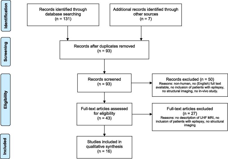
A keyword search strategy was submitted to Pubmed, EMBASE, Cochrane Database and clinicaltrials.gov to select studies on UHF MRI in patients with epilepsy. Follow-up study selection and data extraction were performed following PRISMA guidelines. We focused on I) diagnostic gain of UHF- over conventional MRI, II) concordance of MRI-detected lesion, seizure onset zone and surgical decision-making, and III) postoperative histopathological diagnosis and seizure outcome.
Sixteen observational cohort studies, all using 7T MRI were included. Diagnostic gain of 7T over conventional MRI ranged from 8% to 67%, with a pooled gain of 31%. Novel techniques to visualize pathological processes in epilepsy and lesion detection are discussed. Seizure freedom was achieved in 73% of operated patients; no seizure outcome comparison was made between 7T MRI positive, 7T negative and 3T positive patients. 7T could influence surgical decision-making, with high concordance of lesion and seizure onset zone. Focal cortical dysplasia (54%), hippocampal sclerosis (12%) and gliosis (8.1%) were the most frequently diagnosed histopathological entities.
UHF MRI increases, yet variably, the sensitivity to detect an epileptogenic lesion, showing potential for use in clinical practice. It remains to be established whether this results in improved seizure outcome after surgical treatment. Prospective studies with larger cohorts of epilepsy patients, uniform scan and sequence protocols, and innovative post-processing technology are equally important as further increasing field strengths. Besides technical ameliorations, improved correlation of imaging features with clinical semiology, histopathology and clinical outcome has to be established.
癫痫的致痫灶切除术是一种有循证医学证据支持的治疗药物难治性局灶性癫痫的方法。术前主要的预测指标是磁共振成像(MRI)检测到致痫性病变。应用超高场(UHF)MRI,即场强≥7 特斯拉(T),可能会提高检测到这种病变的敏感性。
我们采用关键词搜索策略,向 Pubmed、EMBASE、Cochrane 数据库和 clinicaltrials.gov 提交了检索词,以筛选关于癫痫患者 UHF MRI 的研究。按照 PRISMA 指南进行了后续的研究选择和数据提取。我们重点关注:I)UHF-MRI 相对于常规 MRI 的诊断增益;II)MRI 检测到的病变、癫痫起始区和手术决策的一致性;III)术后组织病理学诊断和癫痫发作结果。
共纳入了 16 项观察性队列研究,均使用 7T MRI。7T 相对于常规 MRI 的诊断增益范围为 8%至 67%,总体增益为 31%。讨论了用于可视化癫痫病理过程和病变检测的新方法。73%的手术患者达到无癫痫发作;7T MRI 阳性、7T MRI 阴性和 3T MRI 阳性患者之间未进行癫痫发作结果的比较。7T 可能会影响手术决策,病变和癫痫起始区具有高度一致性。最常诊断的组织病理学实体是局灶性皮质发育不良(54%)、海马硬化(12%)和神经胶质增生(8.1%)。
UHF MRI 提高了但变化不定地检测致痫性病变的敏感性,具有在临床实践中应用的潜力。它是否能提高手术治疗后的癫痫发作结果还有待确定。同样重要的是需要进行前瞻性研究,纳入更多的癫痫患者队列、统一的扫描和序列方案、以及创新的后处理技术,同时进一步提高场强。除了技术改进外,还需要建立影像学特征与临床症状学、组织病理学和临床结果的更好相关性。