• 文献检索
  • 文档翻译
  • 深度研究
  • 学术资讯
  • Suppr Zotero 插件Zotero 插件
  • 邀请有礼
  • 套餐&价格
  • 历史记录
应用&插件
Suppr Zotero 插件Zotero 插件浏览器插件Mac 客户端Windows 客户端微信小程序
定价
高级版会员购买积分包购买API积分包
服务
文献检索文档翻译深度研究API 文档MCP 服务
关于我们
关于 Suppr公司介绍联系我们用户协议隐私条款
关注我们

Suppr 超能文献

核心技术专利:CN118964589B侵权必究
粤ICP备2023148730 号-1Suppr @ 2026

文献检索

告别复杂PubMed语法,用中文像聊天一样搜索,搜遍4000万医学文献。AI智能推荐,让科研检索更轻松。

立即免费搜索

文件翻译

保留排版,准确专业,支持PDF/Word/PPT等文件格式,支持 12+语言互译。

免费翻译文档

深度研究

AI帮你快速写综述,25分钟生成高质量综述,智能提取关键信息,辅助科研写作。

立即免费体验

肺部超声中垂直伪像随频率变化的临床影响

Clinical Impact of Vertical Artifacts Changing with Frequency in Lung Ultrasound.

作者信息

Buda Natalia, Skoczylas Agnieszka, Demi Marcello, Wojteczek Anna, Cylwik Jolanta, Soldati Gino

机构信息

Department of Internal Medicine, Connective Tissue Diseases and Geriatric, Medical University of Gdansk, 80-952 Gdansk, Poland.

Department of Geriatrics, National Institute of Geriatrics Rheumatology and Rehabilitation, 02-637 Warsaw, Poland.

出版信息

Diagnostics (Basel). 2021 Feb 26;11(3):401. doi: 10.3390/diagnostics11030401.

DOI:10.3390/diagnostics11030401
PMID:33652906
原文链接:https://pmc.ncbi.nlm.nih.gov/articles/PMC7996921/
Abstract

BACKGROUND

This study concerns the application of lung ultrasound (LUS) for the evaluation of the significance of vertical artifact changes with frequency and pleural line abnormalities in differentiating pulmonary edema from pulmonary fibrosis.

STUDY DESIGN AND METHODS

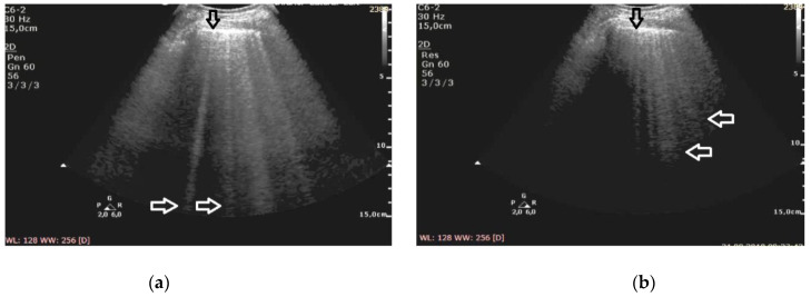
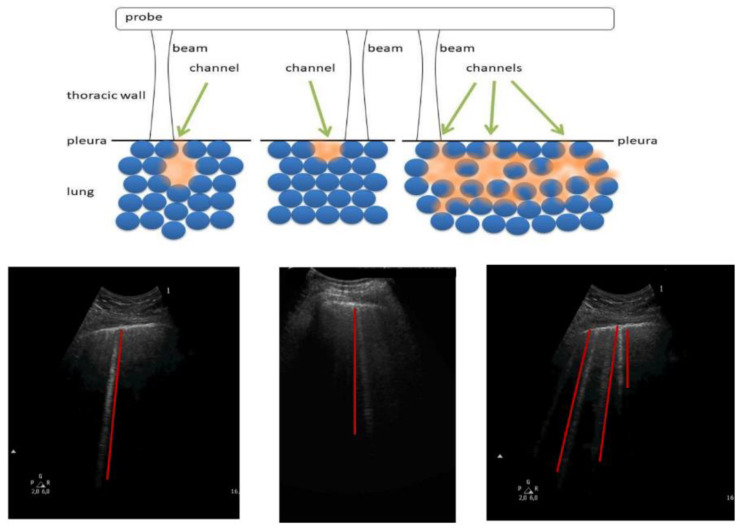
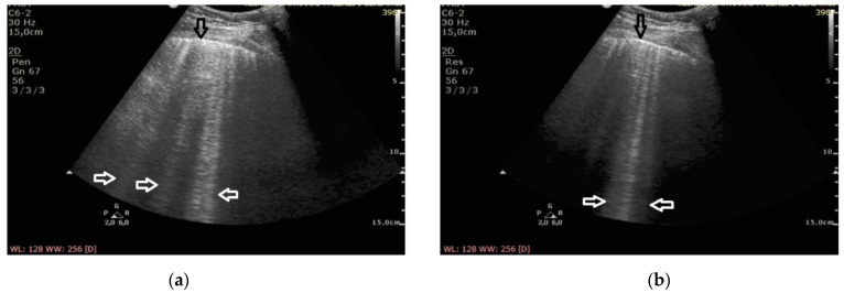
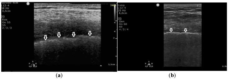
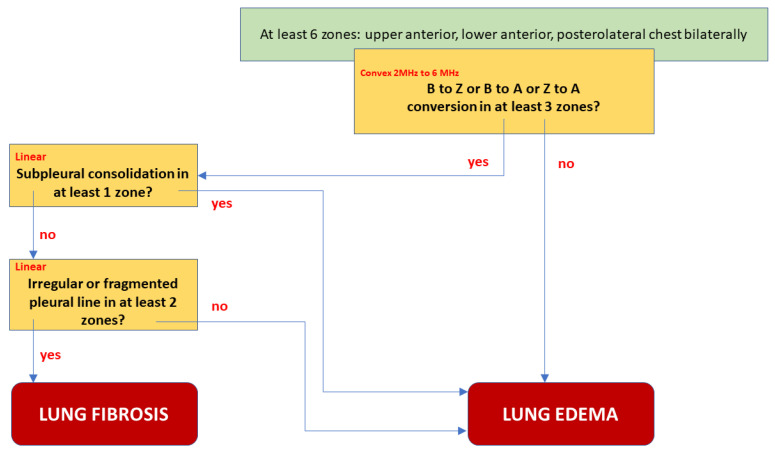
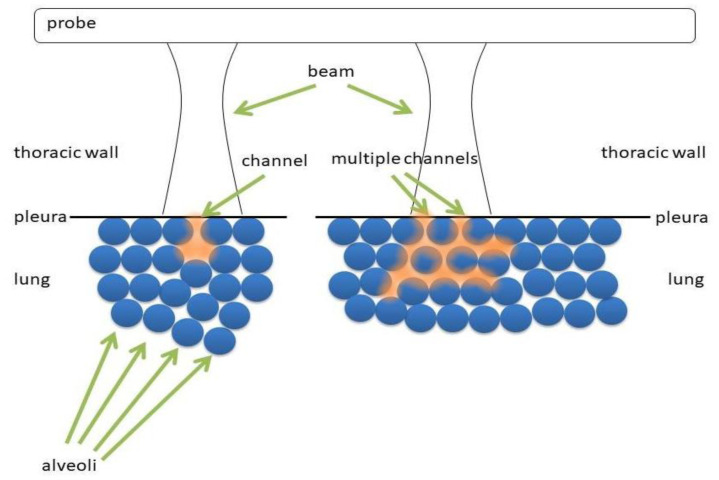
The study was designed as a diagnostic test. Having qualified patients for the study, an ultrasound examination was performed, consistent with a predetermined protocol, and employing convex and linear transducers. We investigated the possibility of B-line artifact conversion depending on the set frequency (2 MHz and 6 MHz), and examined pleural line abnormalities.

RESULTS

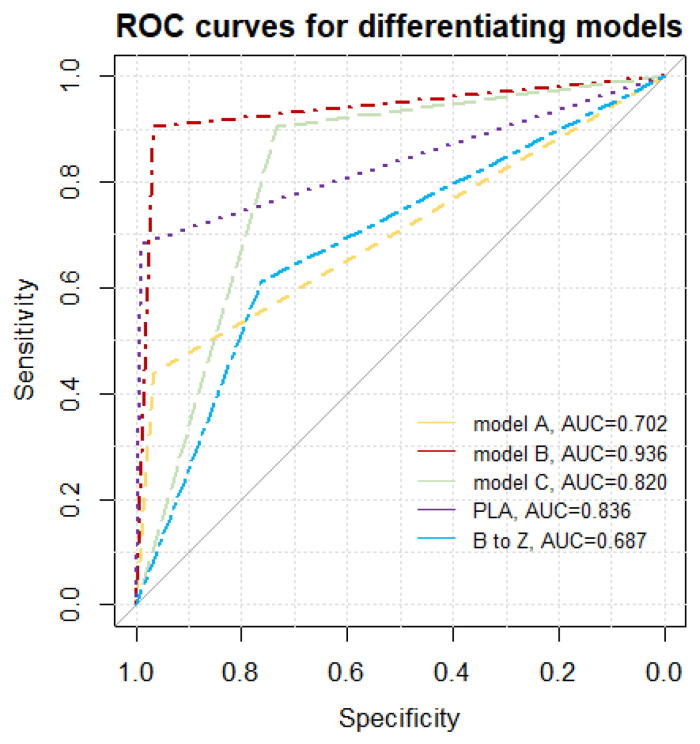
The study group comprised 32 patients with interstitial lung disease (ILD) (and fibrosis) and 30 patients with pulmonary edema. In total, 1941 cineloops were obtained from both groups and analyzed. The employment of both types of transducers (linear and convex) was most effective (specificity 91%, specificity 97%, positive predictive value (PPV) 97%, negative predictive value (NPV) 91%, LR(+) 27,19, LR(-) 0.097, area under curve (AUC) = 0.936, = 7 × 10).

INTERPRETATION

The best accuracy in differentiating the etiology of B-line artifacts was obtained with the use of both types of transducers (linear and convex), complemented with the observation of the conversion of B-line artifacts to Z-line.

摘要

背景

本研究关注肺部超声(LUS)在评估垂直伪像随频率变化的意义以及胸膜线异常在鉴别肺水肿与肺纤维化中的应用。

研究设计与方法

本研究设计为一项诊断试验。在使患者符合研究条件后,按照预定方案进行超声检查,并使用凸阵探头和线阵探头。我们研究了B线伪像根据设定频率(2兆赫和6兆赫)转换的可能性,并检查了胸膜线异常情况。

结果

研究组包括32例间质性肺疾病(ILD)(及纤维化)患者和30例肺水肿患者。两组共获得1941个动态图像并进行分析。使用两种类型的探头(线阵和凸阵)最为有效(敏感度91%,特异度97%,阳性预测值(PPV)97%,阴性预测值(NPV)91%,阳性似然比(LR(+))27.19,阴性似然比(LR(-))0.097,曲线下面积(AUC)=0.936,P = 7×10)。

解读

使用两种类型的探头(线阵和凸阵),并辅以观察B线伪像向Z线的转换,在鉴别B线伪像病因方面可获得最佳准确性。

https://cdn.ncbi.nlm.nih.gov/pmc/blobs/9059/7996921/bf92b8bfc2f0/diagnostics-11-00401-g007.jpg
https://cdn.ncbi.nlm.nih.gov/pmc/blobs/9059/7996921/75e49fb001ad/diagnostics-11-00401-g001.jpg
https://cdn.ncbi.nlm.nih.gov/pmc/blobs/9059/7996921/24c78876def3/diagnostics-11-00401-g002.jpg
https://cdn.ncbi.nlm.nih.gov/pmc/blobs/9059/7996921/4f604ebd70f5/diagnostics-11-00401-g003.jpg
https://cdn.ncbi.nlm.nih.gov/pmc/blobs/9059/7996921/2e09c36a126c/diagnostics-11-00401-g004.jpg
https://cdn.ncbi.nlm.nih.gov/pmc/blobs/9059/7996921/5e0713f1f26e/diagnostics-11-00401-g005.jpg
https://cdn.ncbi.nlm.nih.gov/pmc/blobs/9059/7996921/30f0719da000/diagnostics-11-00401-g006.jpg
https://cdn.ncbi.nlm.nih.gov/pmc/blobs/9059/7996921/bf92b8bfc2f0/diagnostics-11-00401-g007.jpg

相似文献

1
Clinical Impact of Vertical Artifacts Changing with Frequency in Lung Ultrasound.肺部超声中垂直伪像随频率变化的临床影响
Diagnostics (Basel). 2021 Feb 26;11(3):401. doi: 10.3390/diagnostics11030401.
2
[The clinical value of bedside lung ultrasound in the diagnosis of chronic obstructive pulmonary disease and cardiac pulmonary edema].[床旁肺部超声在慢性阻塞性肺疾病及心源性肺水肿诊断中的临床价值]
Zhonghua Wei Zhong Bing Ji Jiu Yi Xue. 2014 Aug;26(8):558-62. doi: 10.3760/cma.j.issn.2095-4352.2014.08.007.
3
Lung Ultrasound in the Screening of Pulmonary Interstitial Involvement Secondary to Systemic Connective Tissue Disease: A Prospective Pilot Study Involving 180 Patients.肺部超声在系统性结缔组织病继发肺间质受累筛查中的应用:一项纳入180例患者的前瞻性初步研究
J Clin Med. 2021 Sep 12;10(18):4114. doi: 10.3390/jcm10184114.
4
Lung Ultrasound Spectroscopy Applied to the Differential Diagnosis of Pulmonary Diseases: An In Vivo Multicenter Clinical Study.肺部超声光谱学在肺部疾病鉴别诊断中的应用:一项体内多中心临床研究。
IEEE Trans Ultrason Ferroelectr Freq Control. 2024 Oct;71(10):1217-1232. doi: 10.1109/TUFFC.2024.3454956. Epub 2024 Oct 10.
5
Integrative Cardiopulmonary Ultrasound for Interstitial Lung Disease Assessment: Correlation between Lung Ultrasound Performance and Cardiac Involvement.用于间质性肺疾病评估的心肺综合超声检查:肺超声表现与心脏受累之间的相关性
Ultrasound Med Biol. 2017 Apr;43(4):744-752. doi: 10.1016/j.ultrasmedbio.2016.11.004. Epub 2017 Jan 7.
6
Lung Ultrasonography in the Evaluation of Interstitial Lung Disease in Systemic Connective Tissue Diseases: Criteria and Severity of Pulmonary Fibrosis - Analysis of 52 Patients.肺部超声在系统性结缔组织病间质性肺疾病评估中的应用:肺纤维化的标准与严重程度——52例患者分析
Ultraschall Med. 2016 Aug;37(4):379-85. doi: 10.1055/s-0041-110590. Epub 2015 Dec 29.
7
Sonographic assessment of interstitial lung disease in patients with rheumatoid arthritis, systemic sclerosis and systemic lupus erythematosus.类风湿关节炎、系统性硬化症和系统性红斑狼疮患者间质性肺疾病的超声评估
Clin Exp Rheumatol. 2015 Jul-Aug;33(4 Suppl 91):S87-91. Epub 2015 Jan 29.
8
Bedside lung ultrasound in the evaluation of acute decompensated heart failure.床旁肺部超声在急性失代偿性心力衰竭评估中的应用
Intern Emerg Med. 2016 Jun;11(4):597-601. doi: 10.1007/s11739-016-1403-0. Epub 2016 Feb 17.
9
Is a linear probe helpful in diagnosing diseases of pulmonary interstitial spaces?线性探头对诊断肺间质疾病有帮助吗?
J Ultrason. 2017 Jun;17(69):136-141. doi: 10.15557/JoU.2017.0021. Epub 2017 Jun 30.
10
Ultrasound mapping of lung changes in idiopathic pulmonary fibrosis.特发性肺纤维化肺部变化的超声成像
Clin Respir J. 2020 Jan;14(1):54-63. doi: 10.1111/crj.13101. Epub 2019 Nov 14.

引用本文的文献

1
Impact of ultrasound settings on lung vertical artefacts: an observational study in mechanically ventilated patients.超声设置对肺部垂直伪像的影响:一项针对机械通气患者的观察性研究
ERJ Open Res. 2025 Jan 13;11(1). doi: 10.1183/23120541.00483-2024. eCollection 2025 Jan.
2
The Role of POCUS to Face COVID-19: A Narrative Review.床旁超声在应对新冠疫情中的作用:一项叙述性综述
J Clin Med. 2024 May 7;13(10):2756. doi: 10.3390/jcm13102756.
3
The Rat Thoracic Ultrasound protocol: scanning technique and normal findings.大鼠胸部超声检查方案:扫描技术及正常表现

本文引用的文献

1
In Vivo Assessment of Pulmonary Fibrosis and Pulmonary Edema in Rodents Using Ultrasound Multiple Scattering.超声多重散射在评估啮齿动物肺纤维化和肺水肿中的应用
IEEE Trans Ultrason Ferroelectr Freq Control. 2020 Nov;67(11):2274-2280. doi: 10.1109/TUFFC.2020.3023611. Epub 2020 Oct 27.
2
Self-Performed Lung Ultrasound for Home Monitoring of a Patient Positive for Coronavirus Disease 2019.自我实施肺部超声用于 2019 冠状病毒病阳性患者的家庭监测。
Chest. 2020 Sep;158(3):e93-e97. doi: 10.1016/j.chest.2020.05.604.
3
Recommendations for Lung Ultrasound in Internal Medicine.
Front Vet Sci. 2024 Feb 19;11:1286614. doi: 10.3389/fvets.2024.1286614. eCollection 2024.
4
Lung Ultrasound: A Leading Diagnostic Tool.肺部超声:一种领先的诊断工具。
Diagnostics (Basel). 2023 May 12;13(10):1710. doi: 10.3390/diagnostics13101710.
5
Lung Ultrasound in the Evaluation of Lung Disease Severity in Children with Clinically Stable Cystic Fibrosis: A Prospective Cross-Sectional Study.肺部超声评估临床稳定型囊性纤维化患儿肺部疾病严重程度:一项前瞻性横断面研究
J Clin Med. 2023 Apr 24;12(9):3086. doi: 10.3390/jcm12093086.
6
The effect of attenuation inside the acoustic traps on the configuration of vertical artifacts in lung ultrasound: an experimental study with simple models.声阱内衰减对肺部超声中垂直伪影形态的影响:采用简单模型的实验研究。
J Med Ultrason (2001). 2022 Oct;49(4):545-553. doi: 10.1007/s10396-022-01244-0. Epub 2022 Aug 5.
7
The Mechanisms Underlying Vertical Artifacts in Lung Ultrasound and Their Proper Utilization for the Evaluation of Cardiogenic Pulmonary Edema.肺部超声垂直伪像的潜在机制及其在评估心源性肺水肿中的合理应用
Diagnostics (Basel). 2022 Jan 20;12(2):252. doi: 10.3390/diagnostics12020252.
8
Effects of positive end-expiratory pressure on lung ultrasound patterns and their correlation with intracranial pressure in mechanically ventilated brain injured patients.正压呼气末对机械通气脑损伤患者肺部超声模式的影响及其与颅内压的相关性。
Crit Care. 2022 Jan 28;26(1):31. doi: 10.1186/s13054-022-03903-7.
9
Vertical Artifacts in Lung Ultrasonography: Some Common Clinician Questions and the Related Engineer Answers.肺部超声检查中的垂直伪像:临床医生的一些常见问题及工程师的相关解答
Diagnostics (Basel). 2022 Jan 16;12(1):215. doi: 10.3390/diagnostics12010215.
10
High-Resolution Computed Tomography and Lung Ultrasound in Patients with Systemic Sclerosis: Which One to Choose?系统性硬化症患者的高分辨率计算机断层扫描和肺部超声:该如何选择?
Diagnostics (Basel). 2021 Dec 7;11(12):2293. doi: 10.3390/diagnostics11122293.
内科肺部超声检查建议
Diagnostics (Basel). 2020 Aug 16;10(8):597. doi: 10.3390/diagnostics10080597.
4
Lung Ultrasound in a Patient With ARDS Secondary to Pancreatitis.胰腺炎继发急性呼吸窘迫综合征患者的肺部超声检查
Chest. 2020 Aug;158(2):e85-e87. doi: 10.1016/j.chest.2019.09.045.
5
Consensus of the Study Group for Point-of-Care Lung Ultrasound in the intensive care management of COVID-19 patients.新冠病毒疾病(COVID-19)患者重症监护管理中床旁肺部超声研究小组共识
Anaesthesiol Intensive Ther. 2020;52(2):83-90. doi: 10.5114/ait.2020.96560.
6
Effect of Different Probes and Expertise on the Interpretation Reliability of Point-of-Care Lung Ultrasound.不同探头和专业水平对即时肺部超声解读可靠性的影响。
Chest. 2020 Apr;157(4):924-931. doi: 10.1016/j.chest.2019.11.013. Epub 2019 Nov 27.
7
Physical Mechanisms Providing Clinical Information From Ultrasound Lung Images: Hypotheses and Early Confirmations.超声肺部图像提供临床信息的物理机制:假说与早期证实。
IEEE Trans Ultrason Ferroelectr Freq Control. 2020 Mar;67(3):612-623. doi: 10.1109/TUFFC.2019.2949597. Epub 2019 Oct 25.
8
Utility of lung ultrasound in ANCA-associated vasculitis with lung involvement.肺超声在伴肺受累的抗中性粒细胞胞质抗体相关性血管炎中的应用。
PLoS One. 2019 Sep 10;14(9):e0222189. doi: 10.1371/journal.pone.0222189. eCollection 2019.
9
Localizing B-Lines in Lung Ultrasonography by Weakly Supervised Deep Learning, In-Vivo Results.通过弱监督深度学习在肺部超声中定位B线:体内结果
IEEE J Biomed Health Inform. 2020 Apr;24(4):957-964. doi: 10.1109/JBHI.2019.2936151. Epub 2019 Aug 19.
10
Thoracic ultrasonography: a narrative review.胸腔超声:叙述性综述。
Intensive Care Med. 2019 Sep;45(9):1200-1211. doi: 10.1007/s00134-019-05725-8. Epub 2019 Aug 15.