• 文献检索
  • 文档翻译
  • 深度研究
  • 学术资讯
  • Suppr Zotero 插件Zotero 插件
  • 邀请有礼
  • 套餐&价格
  • 历史记录
应用&插件
Suppr Zotero 插件Zotero 插件浏览器插件Mac 客户端Windows 客户端微信小程序
定价
高级版会员购买积分包购买API积分包
服务
文献检索文档翻译深度研究API 文档MCP 服务
关于我们
关于 Suppr公司介绍联系我们用户协议隐私条款
关注我们

Suppr 超能文献

核心技术专利:CN118964589B侵权必究
粤ICP备2023148730 号-1Suppr @ 2026

文献检索

告别复杂PubMed语法,用中文像聊天一样搜索,搜遍4000万医学文献。AI智能推荐,让科研检索更轻松。

立即免费搜索

文件翻译

保留排版,准确专业,支持PDF/Word/PPT等文件格式,支持 12+语言互译。

免费翻译文档

深度研究

AI帮你快速写综述,25分钟生成高质量综述,智能提取关键信息,辅助科研写作。

立即免费体验

在细胞形状和膜出芽的决定中,弯曲膜纳米域、被动和主动骨架力的作用。

On the Role of Curved Membrane Nanodomains, and Passive and Active Skeleton Forces in the Determination of Cell Shape and Membrane Budding.

机构信息

Faculty of Electrical Engineering, University of Ljubljana, SI-1000 Ljubljana, Slovenia.

Faculty of Health Sciences, University of Ljubljana, SI-1000 Ljubljana, Slovenia.

出版信息

Int J Mol Sci. 2021 Feb 26;22(5):2348. doi: 10.3390/ijms22052348.

DOI:10.3390/ijms22052348
PMID:33652934
原文链接:https://pmc.ncbi.nlm.nih.gov/articles/PMC7956631/
Abstract

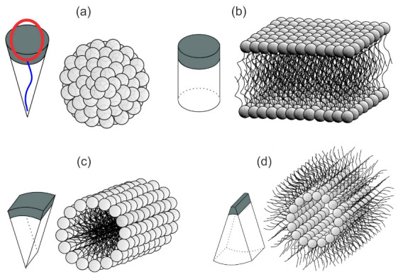
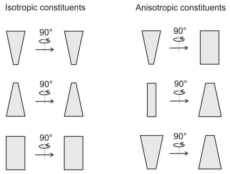
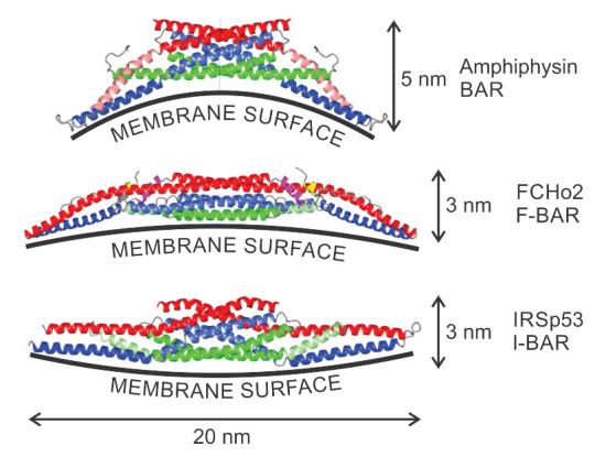
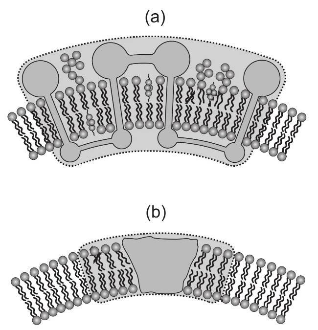
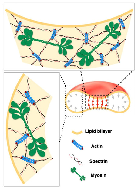
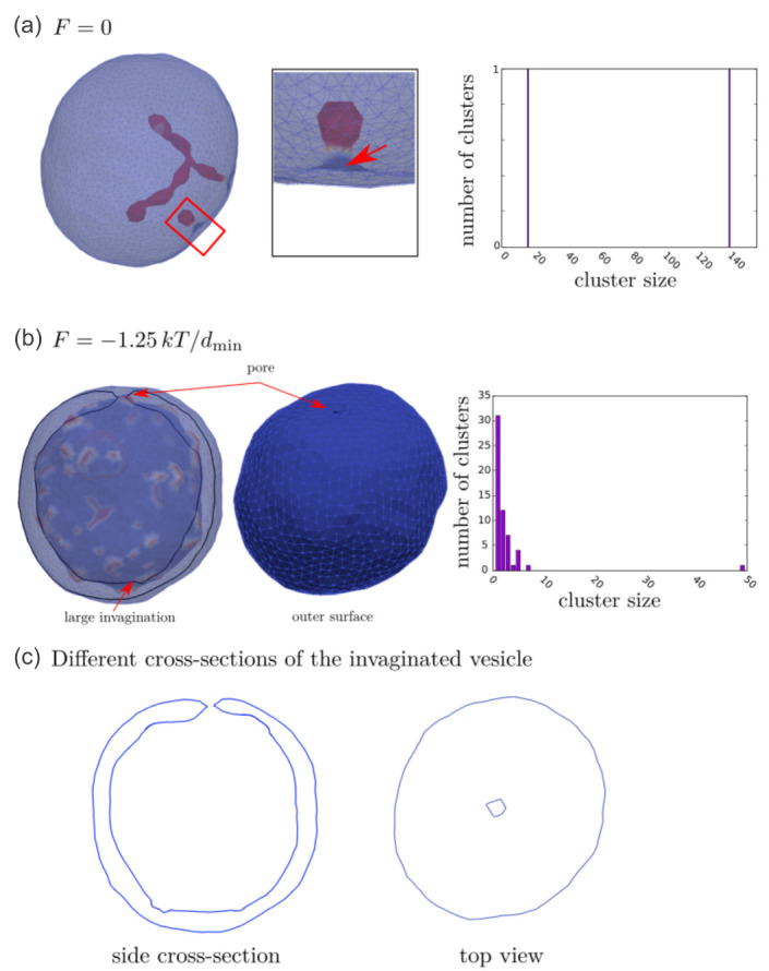
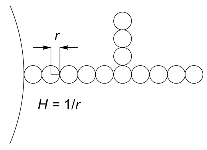
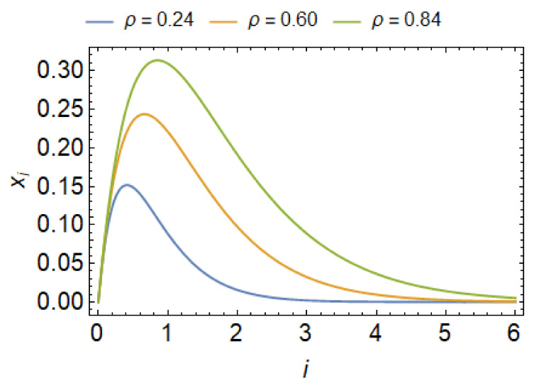
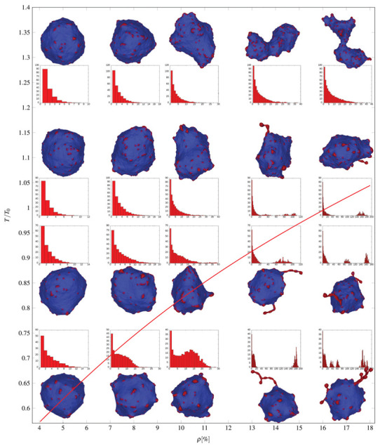
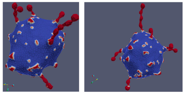
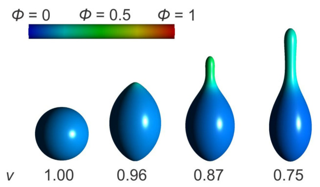
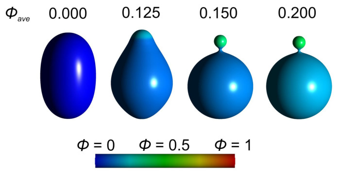
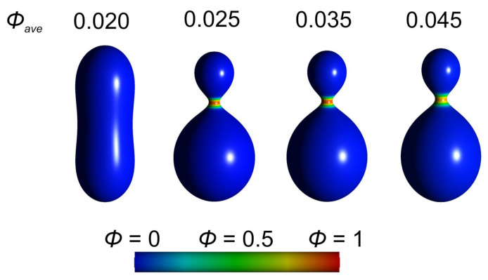
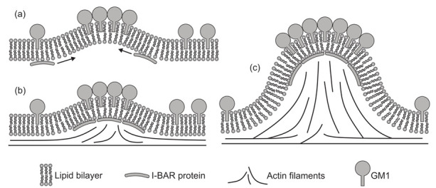
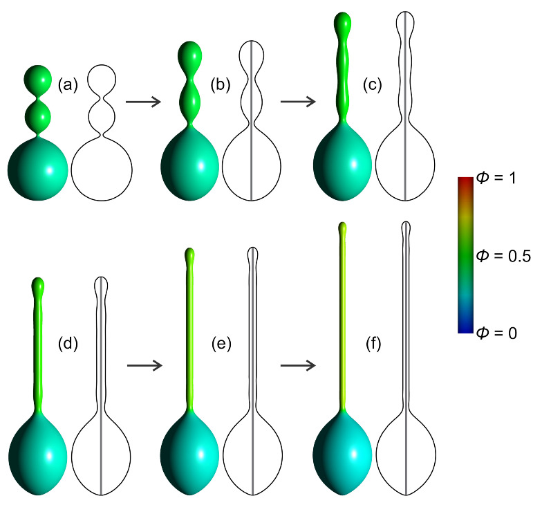
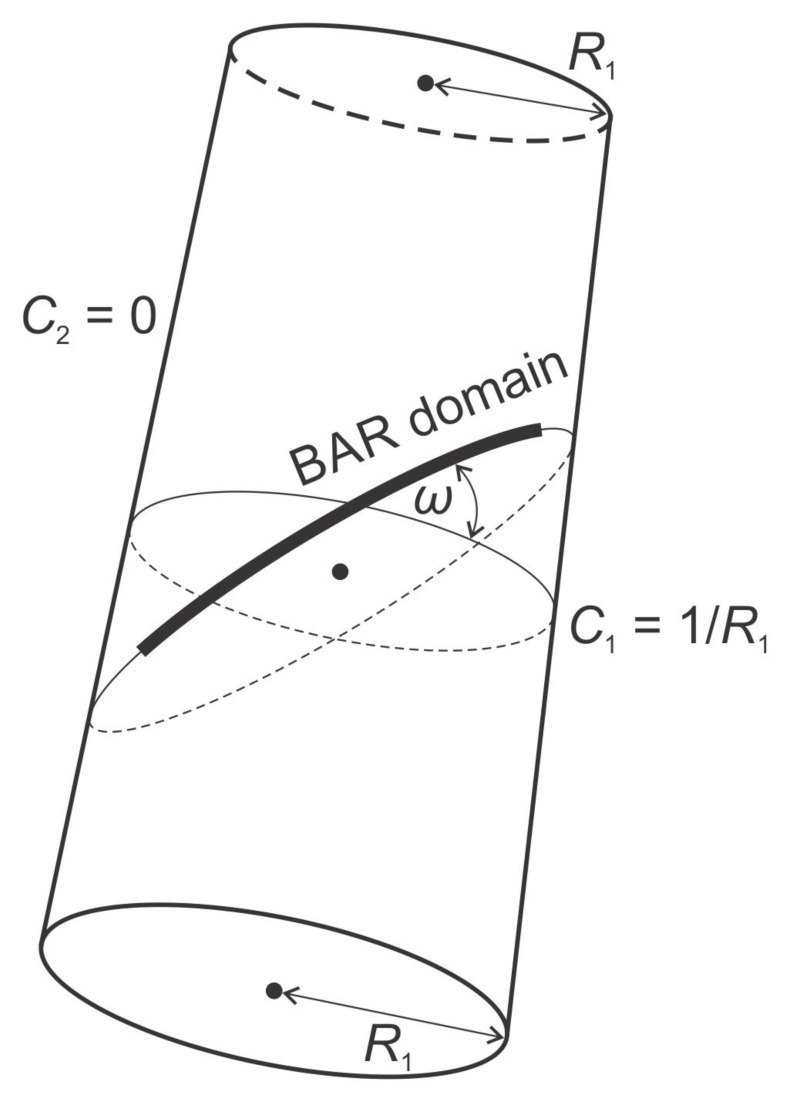
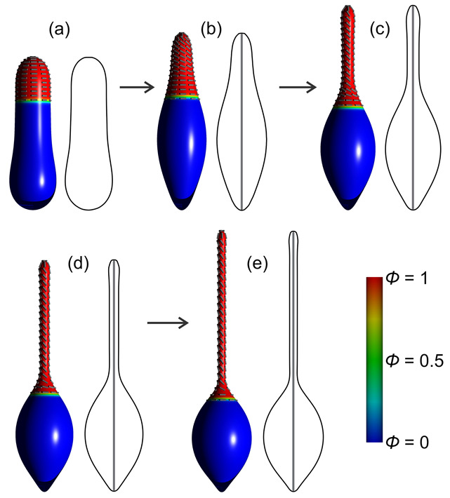
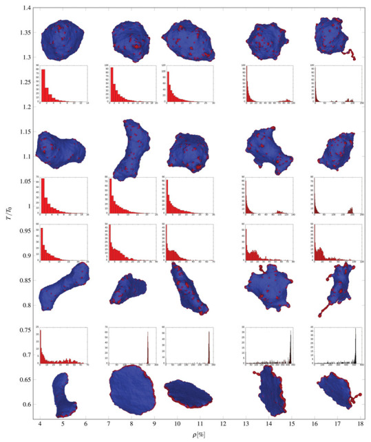
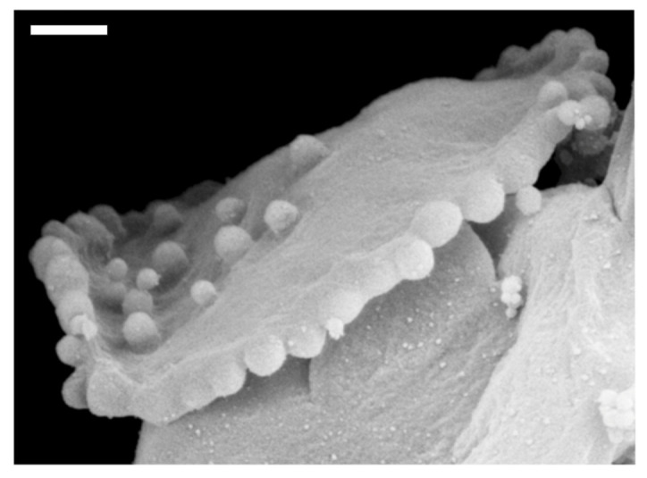
Biological membranes are composed of isotropic and anisotropic curved nanodomains. Anisotropic membrane components, such as Bin/Amphiphysin/Rvs (BAR) superfamily protein domains, could trigger/facilitate the growth of membrane tubular protrusions, while isotropic curved nanodomains may induce undulated (necklace-like) membrane protrusions. We review the role of isotropic and anisotropic membrane nanodomains in stability of tubular and undulated membrane structures generated or stabilized by cyto- or membrane-skeleton. We also describe the theory of spontaneous self-assembly of isotropic curved membrane nanodomains and derive the critical concentration above which the spontaneous necklace-like membrane protrusion growth is favorable. We show that the actin cytoskeleton growth inside the vesicle or cell can change its equilibrium shape, induce higher degree of segregation of membrane nanodomains or even alter the average orientation angle of anisotropic nanodomains such as BAR domains. These effects may indicate whether the actin cytoskeleton role is only to stabilize membrane protrusions or to generate them by stretching the vesicle membrane. Furthermore, we demonstrate that by taking into account the in-plane orientational ordering of anisotropic membrane nanodomains, direct interactions between them and the extrinsic (deviatoric) curvature elasticity, it is possible to explain the experimentally observed stability of oblate (discocyte) shapes of red blood cells in a broad interval of cell reduced volume. Finally, we present results of numerical calculations and Monte-Carlo simulations which indicate that the active forces of membrane skeleton and cytoskeleton applied to plasma membrane may considerably influence cell shape and membrane budding.

摘要

生物膜由各向同性和各向异性的弯曲纳米域组成。各向异性的膜成分,如 Bin/Amphiphysin/Rvs (BAR) 超家族蛋白域,可能触发/促进膜管状突起的生长,而各向同性弯曲纳米域可能诱导起皱(珠串状)的膜突起。我们回顾了各向同性和各向异性膜纳米域在由细胞骨架或膜骨架生成或稳定的管状和起皱膜结构稳定性中的作用。我们还描述了各向同性弯曲膜纳米域自发自组装的理论,并推导出了自发珠串状膜突起生长有利的临界浓度。我们表明,囊泡或细胞内肌动蛋白细胞骨架的生长可以改变其平衡形状,诱导膜纳米域更高程度的分离,甚至改变 BAR 域等各向异性纳米域的平均取向角。这些效应可能表明肌动蛋白细胞骨架的作用仅是稳定膜突起还是通过拉伸囊泡膜来生成它们。此外,我们证明,通过考虑各向异性膜纳米域的面内取向有序性、它们之间的直接相互作用以及外在(偏量)曲率弹性,可以解释在广泛的细胞体积减小区间内观察到的红细胞扁形(盘状)形状的稳定性。最后,我们呈现了数值计算和蒙特卡罗模拟的结果,表明施加于质膜的膜骨架和细胞骨架的主动力可能会显著影响细胞形状和膜出芽。

https://cdn.ncbi.nlm.nih.gov/pmc/blobs/6855/7956631/2efc6ef4b60e/ijms-22-02348-g028.jpg
https://cdn.ncbi.nlm.nih.gov/pmc/blobs/6855/7956631/672dd5435367/ijms-22-02348-g001.jpg
https://cdn.ncbi.nlm.nih.gov/pmc/blobs/6855/7956631/c1564fdce2ab/ijms-22-02348-g002.jpg
https://cdn.ncbi.nlm.nih.gov/pmc/blobs/6855/7956631/4f8c8f351c25/ijms-22-02348-g003.jpg
https://cdn.ncbi.nlm.nih.gov/pmc/blobs/6855/7956631/1cd1263a94b5/ijms-22-02348-g004.jpg
https://cdn.ncbi.nlm.nih.gov/pmc/blobs/6855/7956631/53a19c22864c/ijms-22-02348-g005.jpg
https://cdn.ncbi.nlm.nih.gov/pmc/blobs/6855/7956631/c906142074d5/ijms-22-02348-g006.jpg
https://cdn.ncbi.nlm.nih.gov/pmc/blobs/6855/7956631/0cb847014298/ijms-22-02348-g007.jpg
https://cdn.ncbi.nlm.nih.gov/pmc/blobs/6855/7956631/381ef884878d/ijms-22-02348-g008.jpg
https://cdn.ncbi.nlm.nih.gov/pmc/blobs/6855/7956631/fc88bf28b441/ijms-22-02348-g009.jpg
https://cdn.ncbi.nlm.nih.gov/pmc/blobs/6855/7956631/98b96f86c348/ijms-22-02348-g010.jpg
https://cdn.ncbi.nlm.nih.gov/pmc/blobs/6855/7956631/41ffc6c087ff/ijms-22-02348-g011.jpg
https://cdn.ncbi.nlm.nih.gov/pmc/blobs/6855/7956631/8207f4cea00a/ijms-22-02348-g012.jpg
https://cdn.ncbi.nlm.nih.gov/pmc/blobs/6855/7956631/abe9e662d9aa/ijms-22-02348-g013.jpg
https://cdn.ncbi.nlm.nih.gov/pmc/blobs/6855/7956631/cdd70a372274/ijms-22-02348-g014.jpg
https://cdn.ncbi.nlm.nih.gov/pmc/blobs/6855/7956631/a5bc9ae0effc/ijms-22-02348-g015.jpg
https://cdn.ncbi.nlm.nih.gov/pmc/blobs/6855/7956631/afc1626a4a6e/ijms-22-02348-g016.jpg
https://cdn.ncbi.nlm.nih.gov/pmc/blobs/6855/7956631/faed4e77c510/ijms-22-02348-g017.jpg
https://cdn.ncbi.nlm.nih.gov/pmc/blobs/6855/7956631/491567809a64/ijms-22-02348-g018.jpg
https://cdn.ncbi.nlm.nih.gov/pmc/blobs/6855/7956631/71ee6b10e474/ijms-22-02348-g019.jpg
https://cdn.ncbi.nlm.nih.gov/pmc/blobs/6855/7956631/def76aabd5b7/ijms-22-02348-g020.jpg
https://cdn.ncbi.nlm.nih.gov/pmc/blobs/6855/7956631/9a0361280fb6/ijms-22-02348-g021.jpg
https://cdn.ncbi.nlm.nih.gov/pmc/blobs/6855/7956631/2b3219f2bb83/ijms-22-02348-g022.jpg
https://cdn.ncbi.nlm.nih.gov/pmc/blobs/6855/7956631/a8f74efec75f/ijms-22-02348-g023.jpg
https://cdn.ncbi.nlm.nih.gov/pmc/blobs/6855/7956631/39dfe76cb9f3/ijms-22-02348-g024.jpg
https://cdn.ncbi.nlm.nih.gov/pmc/blobs/6855/7956631/54b486df9dff/ijms-22-02348-g025.jpg
https://cdn.ncbi.nlm.nih.gov/pmc/blobs/6855/7956631/53b5951bada6/ijms-22-02348-g026.jpg
https://cdn.ncbi.nlm.nih.gov/pmc/blobs/6855/7956631/fc5ad9ed6b17/ijms-22-02348-g027.jpg
https://cdn.ncbi.nlm.nih.gov/pmc/blobs/6855/7956631/2efc6ef4b60e/ijms-22-02348-g028.jpg

相似文献

1
On the Role of Curved Membrane Nanodomains, and Passive and Active Skeleton Forces in the Determination of Cell Shape and Membrane Budding.在细胞形状和膜出芽的决定中,弯曲膜纳米域、被动和主动骨架力的作用。
Int J Mol Sci. 2021 Feb 26;22(5):2348. doi: 10.3390/ijms22052348.
2
On the role of external force of actin filaments in the formation of tubular protrusions of closed membrane shapes with anisotropic membrane components.关于肌动蛋白丝的外力在具有各向异性膜成分的封闭膜形状管状突起形成中的作用。
Eur Biophys J. 2017 Dec;46(8):705-718. doi: 10.1007/s00249-017-1212-z. Epub 2017 May 9.
3
Closed membrane shapes with attached BAR domains subject to external force of actin filaments.具有附着BAR结构域的封闭膜形状受到肌动蛋白丝外力作用。
Colloids Surf B Biointerfaces. 2016 May 1;141:132-140. doi: 10.1016/j.colsurfb.2016.01.010. Epub 2016 Jan 21.
4
Possible role of flexible red blood cell membrane nanodomains in the growth and stability of membrane nanotubes.柔性红细胞膜纳米结构域在膜纳米管生长和稳定性中的可能作用。
Blood Cells Mol Dis. 2007 Jul-Aug;39(1):14-23. doi: 10.1016/j.bcmd.2007.02.013. Epub 2007 May 1.
5
Myosin IIA interacts with the spectrin-actin membrane skeleton to control red blood cell membrane curvature and deformability.肌球蛋白 IIA 与血影蛋白-肌动蛋白膜骨架相互作用,以控制红细胞膜的曲率和变形性。
Proc Natl Acad Sci U S A. 2018 May 8;115(19):E4377-E4385. doi: 10.1073/pnas.1718285115. Epub 2018 Apr 2.
6
On the role of membrane anisotropy and BAR proteins in the stability of tubular membrane structures.关于膜各向异性和 BAR 蛋白在管状膜结构稳定性中的作用。
J Biomech. 2012 Jan 10;45(2):231-8. doi: 10.1016/j.jbiomech.2011.10.039. Epub 2011 Dec 3.
7
Theoretical study of vesicle shapes driven by coupling curved proteins and active cytoskeletal forces.由弯曲蛋白与活性细胞骨架力耦合驱动的囊泡形状的理论研究
Soft Matter. 2019 Jul 14;15(26):5319-5330. doi: 10.1039/c8sm02356e. Epub 2019 Jun 25.
8
Active Forces of Myosin Motors May Control Endovesiculation of Red Blood Cells.肌球蛋白马达的活性力可能控制红细胞的内胞吐作用。
Acta Chim Slov. 2020 Jun;67(2):674-681.
9
On the role of anisotropy of membrane components in formation and stabilization of tubular structures in multicomponent membranes.论膜成分各向异性在多组分膜中管状结构形成和稳定中的作用。
PLoS One. 2013 Sep 16;8(9):e73941. doi: 10.1371/journal.pone.0073941. eCollection 2013.
10
Theoretical model of membrane protrusions driven by curved active proteins.由弯曲活性蛋白驱动的膜突出的理论模型。
Front Mol Biosci. 2023 May 9;10:1153420. doi: 10.3389/fmolb.2023.1153420. eCollection 2023.

引用本文的文献

1
Prominosomes - a particular class of extracellular vesicles containing prominin-1/CD133?突起小体——一类含有prominin-1/CD133的特殊细胞外囊泡?
J Nanobiotechnology. 2025 Jan 29;23(1):61. doi: 10.1186/s12951-025-03102-w.
2
Controlled node growth on the surface of polymersomes.聚合物囊泡表面的可控节点生长。
Chem Sci. 2024 Feb 16;15(12):4396-4402. doi: 10.1039/d3sc05915d. eCollection 2024 Mar 20.
3
A minimal physical model for curvotaxis driven by curved protein complexes at the cell's leading edge.细胞前缘弯曲蛋白复合物驱动的趋曲率的最小物理模型。

本文引用的文献

1
Membrane shape remodeling by protein crowding.蛋白质拥挤引起的膜形状重塑
Biophys J. 2021 Jun 15;120(12):2482-2489. doi: 10.1016/j.bpj.2021.04.029. Epub 2021 May 21.
2
Active Forces of Myosin Motors May Control Endovesiculation of Red Blood Cells.肌球蛋白马达的活性力可能控制红细胞的内胞吐作用。
Acta Chim Slov. 2020 Jun;67(2):674-681.
3
Minimizing isotropic and deviatoric membrane energy - An unifying formation mechanism of different cellular membrane nanovesicle types.最小化各向同性和偏量膜能量——不同细胞膜纳米囊泡类型的统一形成机制。
Proc Natl Acad Sci U S A. 2024 Mar 19;121(12):e2306818121. doi: 10.1073/pnas.2306818121. Epub 2024 Mar 15.
4
Molecular Sensing and Manipulation of Protein Oligomerization in Membrane Nanotubes with Bolaamphiphilic Foldamers.利用双两亲螺旋折叠体对膜纳米管中蛋白质寡聚化的分子感应和操控。
J Am Chem Soc. 2023 Nov 22;145(46):25150-25159. doi: 10.1021/jacs.3c05753. Epub 2023 Nov 10.
5
Investigation of nano- and microdomains formed by ceramide 1 phosphate in lipid bilayers.研究神经酰胺 1 磷酸在脂质双层中形成的纳米和微域。
Sci Rep. 2023 Oct 30;13(1):18570. doi: 10.1038/s41598-023-45575-5.
6
Molecular Relay Stations in Membrane Nanotubes: IRSp53 Involved in Actin-Based Force Generation.膜纳米管中的分子接力站:IRSp53 参与基于肌动蛋白的力生成。
Int J Mol Sci. 2023 Aug 23;24(17):13112. doi: 10.3390/ijms241713112.
7
A theoretical model of efficient phagocytosis driven by curved membrane proteins and active cytoskeleton forces.由弯曲膜蛋白和活性细胞骨架力驱动的高效吞噬作用的理论模型。
Soft Matter. 2022 Dec 21;19(1):31-43. doi: 10.1039/d2sm01152b.
8
GTP-stimulated membrane fission by the N-BAR protein AMPH-1.GTP 刺激的 N-BAR 蛋白 AMPH-1 介导的膜裂变。
Traffic. 2023 Jan;24(1):34-47. doi: 10.1111/tra.12875. Epub 2022 Dec 13.
9
Stability of Erythrocyte-Derived Nanovesicles Assessed by Light Scattering and Electron Microscopy.通过光散射和电子显微镜评估红细胞衍生的纳米囊泡的稳定性。
Int J Mol Sci. 2021 Nov 25;22(23):12772. doi: 10.3390/ijms222312772.
10
On the Role of Electrostatic Repulsion in Topological Defect-Driven Membrane Fission.静电排斥在拓扑缺陷驱动的膜裂变中的作用
Membranes (Basel). 2021 Oct 25;11(11):812. doi: 10.3390/membranes11110812.
PLoS One. 2020 Dec 31;15(12):e0244796. doi: 10.1371/journal.pone.0244796. eCollection 2020.
4
Lipid-Composition-Mediated Forces Can Stabilize Tubular Assemblies of I-BAR Proteins.脂质组成介导的力可以稳定 I-BAR 蛋白的管状组装。
Biophys J. 2021 Jan 5;120(1):46-54. doi: 10.1016/j.bpj.2020.11.019. Epub 2020 Nov 26.
5
Non-uniform distribution of myosin-mediated forces governs red blood cell membrane curvature through tension modulation.肌球蛋白介导的力的非均匀分布通过张力调节控制红细胞膜的曲率。
PLoS Comput Biol. 2020 May 26;16(5):e1007890. doi: 10.1371/journal.pcbi.1007890. eCollection 2020 May.
6
Cell-cell junctions as sensors and transducers of mechanical forces.细胞-细胞连接作为机械力的感受器和转换器。
Biochim Biophys Acta Biomembr. 2020 Sep 1;1862(9):183316. doi: 10.1016/j.bbamem.2020.183316. Epub 2020 Apr 28.
7
Normal red blood cells' shape stabilized by membrane's in-plane ordering.正常的红细胞形状由膜的面内有序性稳定。
Sci Rep. 2019 Dec 24;9(1):19742. doi: 10.1038/s41598-019-56128-0.
8
Cell confinement reveals a branched-actin independent circuit for neutrophil polarity.细胞限制揭示了中性粒细胞极性的分支肌动蛋白独立回路。
PLoS Biol. 2019 Oct 10;17(10):e3000457. doi: 10.1371/journal.pbio.3000457. eCollection 2019 Oct.
9
Curving Cells Inside and Out: Roles of BAR Domain Proteins in Membrane Shaping and Its Cellular Implications.内外弯曲的细胞:BAR 结构域蛋白在膜塑形及其细胞意义中的作用。
Annu Rev Cell Dev Biol. 2019 Oct 6;35:111-129. doi: 10.1146/annurev-cellbio-100617-060558. Epub 2019 Jul 23.
10
Theoretical study of vesicle shapes driven by coupling curved proteins and active cytoskeletal forces.由弯曲蛋白与活性细胞骨架力耦合驱动的囊泡形状的理论研究
Soft Matter. 2019 Jul 14;15(26):5319-5330. doi: 10.1039/c8sm02356e. Epub 2019 Jun 25.