Matsuki Mitsuru, Numoto Isao, Hamakawa Takefumi, Ishii Kazunari, Chikugo Takaaki
Department of Radiology, Kindai University Faculty of Medicine, Osakasayama-city, Osaka, Japan.
Department of Pathology, Kindai University Faculty of Medicine, Osakasayama-city, Osaka, Japan.
Gynecol Oncol Rep. 2021 Feb 19;36:100733. doi: 10.1016/j.gore.2021.100733. eCollection 2021 May.
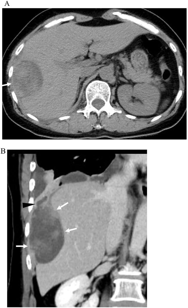
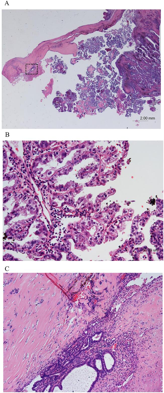
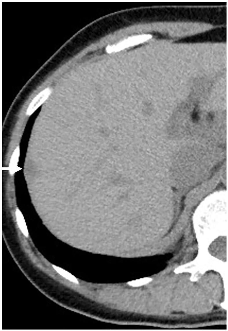
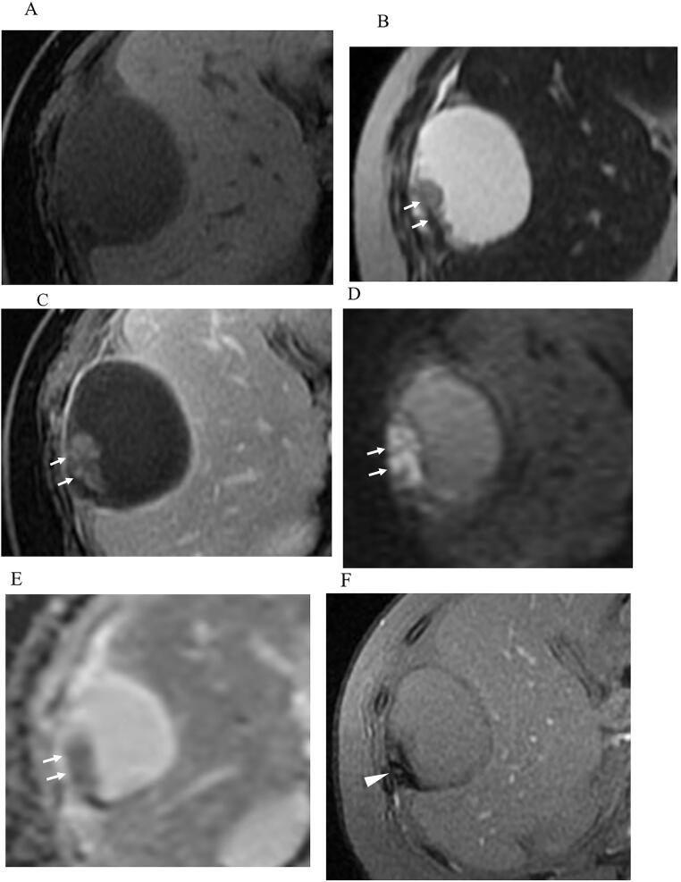
Diaphragmatic endometriosis is extremely rare. Although endometriosis is considered generally benign, malignant transformation of endometriosis was reported in 1925. Multiple studies have since described clear cell carcinoma (CCC) or endometrioid carcinoma arising from ovarian endometriosis. Previously, only two reports of primary diaphragmatic CCC were reported, in which coexistent endometriosis with CCC was not histologically proven. We report a case of a 55-year-old postmenopausal woman who was admitted to Kindai university hospital for the examination of a cystic mass with papillary components in the right diaphragm. On her past medical history, abdominal hysterectomy and bilateral salpingo-oophorectomy was performed for high-grade cervical intraepithelial neoplasia, uterine myoma, and bilateral ovarian endometriosis 5 years ago. Unenhanced CT performed 5 years ago, showed a nodular lesion with low density in the right diaphragm, consistent with diaphragmatic endometriosis. Magnetic resonance imaging during this admission, showed a cystic mass with papillary components in the right diaphragm and a T2*-weighted gradient echo imaging showed partial low signal intensity in the papillary components and cyst wall, which was suspected to represent hemosiderin deposition. Based on these serial images, malignant transformation of diaphragmatic endometriosis was suspected. Under, open abdominal combined resection of the mass and part of the diaphragm was performed. Endometriosis implants were detected on the pelvic peritoneum. Histopathological examination revealed clear cell carcinoma associated with endometriosis and hemosiderin deposition in the cyst wall. T2*-weighted gradient echo imaging was useful in the detection of hemosiderin deposition caused by the coexistent endometriosis. When a cystic mass with papillary components and cyst wall with hemosiderin deposits are encountered on MR images, malignant transformation of endometriosis is suspected and a detailed medical history should be determined and the possibility of concurrent endometriosis or adenomyosis should be investigated, as should the potential existence of diaphragmatic endometriosis in previous images.
膈子宫内膜异位症极为罕见。虽然子宫内膜异位症一般被认为是良性的,但1925年有报道称子宫内膜异位症发生了恶性转化。此后多项研究描述了源于卵巢子宫内膜异位症的透明细胞癌(CCC)或子宫内膜样癌。此前,仅报道过两例原发性膈CCC,其中CCC合并子宫内膜异位症在组织学上未得到证实。我们报告一例55岁绝经后女性,因检查右侧膈肌有带乳头成分的囊性肿块入住近畿大学医院。既往病史显示,5年前因高级别宫颈上皮内瘤变、子宫肌瘤和双侧卵巢子宫内膜异位症行腹式子宫切除术和双侧输卵管卵巢切除术。5年前的平扫CT显示右侧膈肌有一个低密度结节性病变,符合膈子宫内膜异位症。此次入院时的磁共振成像显示右侧膈肌有一个带乳头成分的囊性肿块,T2 *加权梯度回波成像显示乳头成分和囊壁部分低信号强度,怀疑代表含铁血黄素沉积。基于这些系列图像,怀疑膈子宫内膜异位症发生了恶性转化。在开放手术下,对肿块和部分膈肌进行了联合切除。在盆腔腹膜上检测到子宫内膜异位症植入物。组织病理学检查显示为与子宫内膜异位症相关的透明细胞癌以及囊壁中的含铁血黄素沉积。T2 *加权梯度回波成像有助于检测共存子宫内膜异位症引起的含铁血黄素沉积。当在磁共振图像上遇到有乳头成分的囊性肿块和有含铁血黄素沉积的囊壁时,怀疑子宫内膜异位症发生了恶性转化,应确定详细的病史,并调查是否并发子宫内膜异位症或子宫腺肌病,以及既往图像中是否存在膈子宫内膜异位症的可能性。